Our Specialists for Adult Degenerative Scoliosis
Adult deformity surgery is one of the most technically demanding operations in spine. Getting the alignment right the first time matters for decades of your life — which is why it should be done by a team that does it often and uses modern tools to plan and execute the correction.
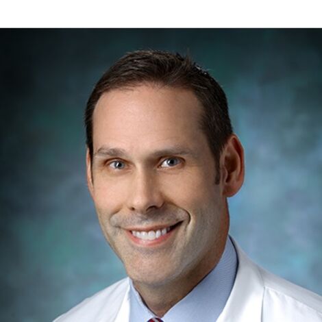
Dr. Witham is Section Chief of Spine Surgery at UChicago and a pioneer in augmented reality-guided spine surgery. In 2020 he performed the first FDA-cleared AR-guided spine surgery on a living patient using the xvision system, and he has published extensively on AR-assisted pedicle screw placement. He came to UChicago from Johns Hopkins, where he was Professor of Neurological Surgery and Orthopaedic Surgery and served as co-program director of the neurosurgery residency. Dr. Witham leads spine surgery at UChicago and is one of the pioneers of augmented reality-guided pedicle screw placement — a technology that matters enormously in deformity cases where 15-20 screws must be placed accurately in rotated, osteoporotic bone. His team reported over 98% screw accuracy in the first 205 consecutive AR-assisted screws (J Neurosurg Spine, 2022).
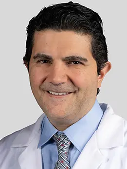
Dr. Bydon is the Chair of Neurological Surgery at UChicago and a pioneer in minimally invasive and robotic spine surgery. He led the stem cell trial, the first-in-human stem cell therapy for spinal cord injury, and holds 12 medical device patents with over 600 peer-reviewed publications. He was recruited to UChicago from Mayo Clinic. Dr. Bydon came to UChicago from Mayo Clinic, where his group published extensively on minimally invasive and lateral interbody approaches to adult degenerative scoliosis and on how to make deformity correction reproducible through data-driven surgical planning. For patients who want the benefits of MIS correction without giving up on alignment targets, he is frequently the surgeon involved.
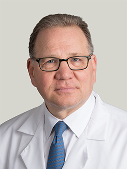
Dr. Herman is Program Director of the Neurological Surgery Residency and a complex spine surgeon who practices the full spectrum of spine and neurorestoration procedures. He co-developed a fully implantable wireless intraspinal microstimulation device for restoring motor function after spinal cord injury, with publications in Artificial Organs and Scientific Reports. He has been named a Top Chicago Doctor for over a decade. Dr. Herman runs a complex spine practice at UChicago and sees adult deformity patients weekly, including patients with prior failed spine surgery and those with combined deformity and instability. For patients whose scoliosis exists alongside other complicated spine pathology, he is one of the surgeons most likely to be involved in planning the operation.
What Is Adult Degenerative Scoliosis?
Adult degenerative scoliosis — sometimes called de novo scoliosis — is a spinal curvature that develops in adulthood, usually after age 50, as a consequence of asymmetric wear-and-tear in your discs and facet joints. It is not the same disease as the scoliosis teenagers get. Adolescent idiopathic scoliosis is primarily a cosmetic problem in a growing spine; adult degenerative scoliosis is a mechanical and neurologic problem in a spine that is breaking down.
As one side of a disc collapses faster than the other, the vertebrae above it tilt. Over years, that tilting stacks up into a curve. The same process narrows the openings where nerves exit the spine, which is why many patients feel the condition first in their legs rather than their back.
Studies of healthy elderly volunteers have found that the majority of people over 60 have some degree of scoliosis if you look hard enough. The important question is not whether you have a curve — many people do — but whether your curve is causing pain, nerve compression, or a loss of sagittal balance (your ability to stand upright without leaning forward).
That last concept is critical, and it is where modern deformity surgery lives. When the lumbar spine loses its natural forward curve (lordosis), your head drifts forward over your pelvis. Your hips, knees, and back muscles have to work constantly to hold you up. Within a few years you feel exhausted after short walks, can't stand at a counter to cook, and begin to lean on a grocery cart. That functional collapse is what we are actually trying to fix.
At a Glance
- Adult degenerative scoliosis is a sideways curve of the spine that develops after skeletal maturity, usually after age 50, as discs and joints wear out asymmetrically
- The problem is often not the curve itself but the loss of forward-backward (sagittal) balance — which is what actually drives pain and disability
- The main symptoms are back pain, leg pain from pinched nerves, and a feeling of pitching forward when you walk
- Surgical goals are measured in degrees and millimeters using specific parameters (PI-LL, SVA, T1 pelvic angle) — not just how straight your x-ray looks
- Modern correction often combines lateral interbody fusion (LLIF) from the side with posterior instrumentation from the back, with navigation or augmented reality to place screws accurately
Have imaging or a diagnosis already?
We'll have a specialist review your MRI and records — often within 24 hours.
What Does It Feel Like?
Adult scoliosis rarely announces itself with a dramatic event. It creeps in over years. Most patients come in describing some combination of the following:
Back pain
- Aching or burning low back pain that is worse with standing and walking, better with sitting or lying down
- Pain that is worst at the end of the day
- A sense that your back is collapsing or giving out when you stand up for too long
Leg pain and nerve symptoms
- Shooting pain, numbness, or tingling down one or both legs (radiculopathy)
- Heaviness, cramping, or weakness in the legs that comes on after walking a block or two and goes away when you sit (neurogenic claudication)
- Occasionally, foot drop or difficulty clearing the toe when walking
Posture and balance changes
- A feeling that you are pitching forward when you walk or stand
- Needing to hold onto a counter, cart, or walker to stay upright
- One shoulder sitting higher than the other, or one hip appearing more prominent
- A rib hump or asymmetry that you first noticed in photos or in a mirror
- Fatigue after short walks — because your back muscles are working overtime to hold you up
Symptoms that are not typical of scoliosis alone, and that should be taken seriously as possible signs of a separate problem, include new bowel or bladder changes, severe night pain, unexplained weight loss, or sudden severe weakness. Those warrant urgent evaluation.
How Is It Diagnosed?
The diagnosis starts with a careful history and physical exam, but imaging is where adult scoliosis gets its real characterization.
Standing full-length x-rays
Unlike most spine problems, scoliosis cannot be properly evaluated on an MRI alone. You need 36-inch standing x-rays of the entire spine from the base of the skull to the femoral heads, taken while you are upright and bearing weight. These films let us measure:
- Cobb angle — the magnitude of the sideways curve, in degrees
- Sagittal vertical axis (SVA) — how far forward your head sits over your pelvis; anything more than about 5 cm is abnormal
- Pelvic incidence minus lumbar lordosis (PI-LL) — essentially a measurement of whether you have enough forward curve in your lower back to match your pelvic anatomy; a mismatch over 10 degrees correlates strongly with disability
- Pelvic tilt (PT) — how much your pelvis has rotated backward to compensate for the loss of lumbar lordosis
- T1 pelvic angle (T1PA) — a newer composite measure that captures both pelvic compensation and trunk forward-lean in one number
These measurements are not academic. They are the numbers your surgeon uses to plan exactly how much correction you need and where. The SRS-Schwab classification groups patients by curve type and by the severity of each of these sagittal modifiers, and the classification correlates tightly with disability.
MRI
An MRI shows us the soft tissue — discs, nerves, ligaments, and any spinal stenosis contributing to leg symptoms. This tells us which levels need to be decompressed in addition to being realigned and fused.
CT scan
A CT scan gives the best view of the bone — including old fractures, bone quality, and the exact anatomy of the pedicles where screws will be placed. In patients with osteoporosis, a DEXA scan is also essential before any major deformity surgery, because poor bone quality dramatically raises the risk of screws pulling out and of fractures at the top of the construct.
Types of Adult Scoliosis
Not every adult with a curved spine has the same disease, and the distinction actually matters for how we treat you.
De novo degenerative scoliosis
This is the most common type we treat. The curve develops in adulthood, typically after age 50, from asymmetric degeneration of discs and facet joints. Curves are usually in the lumbar spine, tend to be shorter (5-8 vertebrae), and are strongly associated with spinal stenosis and leg symptoms. The Cobb angle is often modest (20-40 degrees), but the sagittal imbalance and nerve compression can be severe.
Progressive idiopathic scoliosis
These patients had scoliosis as adolescents — sometimes known, sometimes not — and the curve has continued to progress into adulthood. Curves tend to be larger (often over 40-50 degrees), longer, and involve the thoracic spine as well as the lumbar. Back pain dominates early, with leg symptoms appearing later as the curve collapses further.
How the SRS-Schwab classification groups all of this
The SRS-Schwab adult spinal deformity classification does not care whether your curve is de novo or progressive. It groups you by:
- Curve type — thoracic only, thoracolumbar/lumbar, double, or sagittal-plane only (no coronal curve at all)
- PI-LL mismatch — 0 (normal), + (moderate), or ++ (marked)
- Sagittal vertical axis — 0, +, or ++
- Pelvic tilt — 0, +, or ++
A patient classified as L,++,++,++ has a lumbar curve with severe sagittal imbalance in every parameter and is far more disabled — and needs far more aggressive correction — than a patient classified as T,0,0,0. This framework is what lets your surgeon explain, in concrete terms, why you need the operation you need.
How Is It Treated?
Nonoperative care comes first
Most patients with adult degenerative scoliosis do not need surgery. The first line of treatment is nonoperative: physical therapy focused on core and hip strength, anti-inflammatory medication, targeted epidural steroid injections for radicular leg pain, and, in selected cases, bracing. Many patients live well for years with this approach.
Where nonoperative care falls short is in patients whose sagittal balance has already collapsed. Injections and PT do not put lordosis back into a flat lumbar spine. For patients with severe PI-LL mismatch, large SVA, and progressive disability despite maximal nonoperative care, surgery is the only treatment that meaningfully changes the trajectory — and prospective multicenter data show that operative patients gain substantial, durable improvements in pain and function while matched nonoperative patients, on average, do not.
What deformity surgery is actually trying to do
Modern adult deformity surgery has three goals, in this order:
- Decompress the nerves that are being pinched, usually in the lumbar spine
- Rebuild sagittal alignment — put lumbar lordosis back into the lower back so your head sits over your pelvis and your muscles can rest
- Stabilize the correction with fusion and instrumentation so it holds for the long term
Targets are individualized and age-adjusted. A 45-year-old needs a different alignment target than a 75-year-old; pushing an elderly patient to a "young" target is actually harmful and drives complications at the top of the construct.
Lateral interbody fusion (LLIF) plus posterior instrumentation
For many adult scoliosis patients, the workhorse operation is a staged or same-day combined approach:
- Lateral lumbar interbody fusion (LLIF) — through a small incision in your side, the surgeon removes the collapsed discs and inserts large, lordotic cages. This restores disc height, opens the nerve openings indirectly, and puts lordosis back into the spine without cutting through the back muscles.
- Posterior instrumentation — pedicle screws and rods placed from the back to lock the correction in and add direct decompression wherever nerves are still pinched.
For patients with more severe, rigid deformity, a more aggressive osteotomy (such as a pedicle subtraction osteotomy) may be required to unlock a stiff spine before it can be realigned.
MIS versus open correction
Minimally invasive techniques — lateral access, percutaneous screws, and tubular decompression — reduce blood loss, transfusion requirements, infection rates, and hospital stay compared with traditional open surgery. For the right curves, MIS or hybrid techniques can achieve comparable alignment correction to open surgery with substantially lower morbidity. For very large, rigid, or three-column deformities, open surgery with formal osteotomies remains the most reliable way to hit alignment targets. Choosing between the two is a case-by-case decision based on your curve, your bone quality, your age, and your overall health.
Navigation and augmented reality
Placing 15-20 pedicle screws accurately in a rotated, deformed spine is technically demanding and was historically one of the main sources of complications. Intraoperative navigation and augmented reality have changed that. Using a head-mounted AR display, the surgeon sees the planned screw trajectory overlaid directly on the patient, reported in a consecutive series of 205 screws with over 98% accuracy. This is now a routine part of deformity surgery at UChicago.
What recovery looks like
Deformity surgery is a big operation. Expect 4-7 days in the hospital, several weeks of limited activity, and 6-12 months to feel the full benefit of the correction. Most patients are walking the day after surgery and are back to most activities of daily living within 6-8 weeks. The biggest single predictor of a good long-term result is whether the alignment target was hit in the operating room — which is why the planning and the execution are as important as the recovery itself.
Considering surgery or planning a second opinion?
Our multidisciplinary team reviews complex cases together. You'll get a coordinated plan, not one opinion.
What Are the Outcomes?
Well-selected adult deformity patients who undergo surgery typically see large, durable improvements in back pain, leg pain, walking tolerance, and quality of life. In prospective multicenter data from the International Spine Study Group (ISSG), operative patients achieve significant improvements in Oswestry Disability Index (ODI) and SRS-22/SF-36 scores at 2 years, while propensity-matched nonoperative patients on average maintain their baseline disability. The flip side of this is that adult deformity surgery has one of the highest complication rates in all of spine — which is why who does the operation matters.
| Outcome | Typical rate | What to know |
|---|---|---|
| Meaningful improvement in back pain at 2 years | ~70-80% | Much higher than matched nonoperative care |
| Meaningful improvement in leg pain at 2 years | ~70% | Driven by decompression + indirect decompression from LLIF |
| Any perioperative complication | ~50% | Includes minor and transient events; most do not affect outcome |
| Major complication by 2 years | ~40% | Has fallen over the last decade as techniques have matured |
| Proximal junctional kyphosis (PJK) | ~25-40% | Radiographic finding; often asymptomatic |
| Proximal junctional failure (PJF) requiring revision | ~5-10% | Higher with osteoporosis, overcorrection, older age |
| Revision surgery by 2 years | ~15-20% | Lower with personalized planning and age-adjusted targets |
Two patterns emerge from these numbers. First, the right operation does work — most appropriately selected patients see substantial, measurable improvement in function and pain. Second, complications are common, and they are strongly related to how well the surgery is planned and executed. Age-adjusted alignment targets, preoperative bone-quality optimization, ligament augmentation at the top of the construct, and accurate screw placement have all been shown to reduce PJK, PJF, and reoperation rates. That is the whole argument for being treated by a team that does a lot of this work.
References
Have Questions About Adult Scoliosis?
Our team is here to review your case, discuss your options, and help you take the next step.
Schedule: (773) 702-2123