Our Specialists for Cerebral Arteriovenous Malformation (AVM)
Brain AVMs are rare, and the decision to treat one is nearly as consequential as the treatment itself. At UChicago, AVM cases are reviewed jointly by cerebrovascular neurosurgeons, endovascular specialists, and radiosurgery experts before a recommendation is ever made.
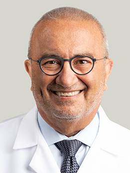
Dr. Awad is Section Chief of Vascular Neurosurgery and a world authority on cerebral cavernous malformations. He discovered the Common Hispanic CCM1 and Ashkenazi Jewish CCM2 mutations and leads the nation's first designated CCM Center of Excellence, with continuous NIH funding since 1998. He has authored more than 400 publications with over 100,000 citations, serves as past President of the Congress of Neurological Surgeons, and is an elected member of the Association of American Physicians. Dr. Awad is the John Harper Seeley Professor and Director of Neurovascular Surgery at UChicago and one of the most cited cerebrovascular surgeons in the world on brain vascular malformations. He has chaired the NIH-funded research consortium on cerebral cavernous malformations for more than a decade and co-authored the AHA/ASA scientific statement on the management of brain AVMs (Stroke, 2017).
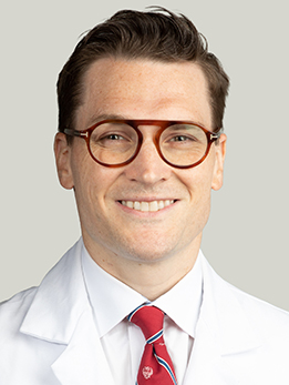
Dr. Polster is Co-Director of the Stroke Center and directs the Skull Base & Neurovascular Laboratory at UChicago. He leads the Gut-Brain Axis Laboratory, where he was the first to demonstrate that the gut microbiome modulates the effects of radiosurgery on the neurovascular unit. His work is funded at the NIH NINDS R-level, and he has published in Nature Communications, Blood, Stroke, and the Journal of Neurosurgery. Dr. Polster is a cerebrovascular neurosurgeon focused on the open microsurgical and multimodal treatment of AVMs, cavernous malformations, and complex aneurysms, and works alongside Dr. Awad on imaging-biomarker research for brain vascular lesions (American Journal of Neuroradiology, 2018).

Dr. Warnke is an international leader in functional neurosurgery and has performed over 6,000 stereotactic surgeries and more than 3,000 brain tumor surgeries. He is only the second neurosurgeon worldwide to perform laser hemispherotomy, and he has completed over 400 laser ablation surgeries since arriving at UChicago. He is funded by four NIH grants including the BRAIN Initiative, and he directs the NAUTILUS trial for thalamic stimulation in drug-resistant epilepsy. For small, deep AVMs in areas where open resection is not the safest choice, Dr. Warnke — Director of Stereotactic and Functional Neurosurgery at UChicago — brings decades of stereotactic planning experience to radiosurgical targeting and the management of residual or recurrent lesions.
What Is a Brain AVM?
A cerebral arteriovenous malformation is a tangle of abnormal blood vessels where arteries connect directly to veins without the normal network of tiny capillaries in between. Capillaries usually act as a buffer, slowing blood down before it enters the veins. Without that buffer, high-pressure arterial blood slams into thin-walled veins — and over years or decades, those veins can weaken, balloon, and rupture.
AVMs are rare. They affect roughly 10 to 18 people per 100,000, and most are congenital, meaning you were born with them. They can sit silently for decades before causing any trouble. When they do cause problems, it's usually because they bleed, trigger a seizure, or show up on a scan ordered for an unrelated headache.
The annual risk of hemorrhage from an untreated AVM averages about 2 to 4% per year, but that number varies significantly based on features of the specific AVM — whether it has bled before, whether its drainage is deep, whether it sits in a critical part of the brain, and whether there are associated aneurysms inside the tangle.
At a Glance
- A brain AVM is a tangle of abnormal arteries and veins that bypasses the normal capillary network, forcing high-pressure blood directly into fragile draining veins
- Most AVMs are found after a hemorrhage, a seizure, or as an incidental finding on a scan done for another reason
- The single most important number is the Spetzler-Martin grade (I through V), which predicts how safely an AVM can be surgically removed
- There are four real options: watchful waiting, microsurgical resection, endovascular embolization, and stereotactic radiosurgery — often in combination
- For unruptured AVMs, the ARUBA trial changed the conversation, and the decision to treat should be made carefully by a team that sees these lesions regularly
Have imaging or a diagnosis already?
We'll have a specialist review your MRI and records — often within 24 hours.
What Does It Feel Like?
Most AVMs cause no symptoms at all until something goes wrong. When symptoms do appear, they usually fall into one of three categories.
Sudden hemorrhage (the most common presentation)
- A sudden, severe headache — often described as the worst headache of your life
- Nausea and vomiting
- Weakness or numbness on one side of the body
- Trouble speaking or understanding speech
- Loss of consciousness or seizure at onset
- Vision changes, depending on the AVM's location
Seizures
- A first-ever seizure in an adult, sometimes without any other warning signs
- Focal seizures (twitching, sensory changes, or brief episodes of confusion) if the AVM sits near the brain's surface
Other, less specific symptoms
- Chronic headaches, sometimes with migraine-like features
- Progressive weakness, numbness, or coordination problems
- A bruit — a rhythmic whooshing sound you can hear inside your head that matches your pulse
A substantial minority of AVMs are found entirely by accident — on an MRI ordered for something else. Those incidental AVMs are the hardest decisions in the field.
How Is It Diagnosed?
If an AVM is suspected after a sudden headache or a new seizure, the workup typically moves quickly.
CT and CT angiography
In the emergency department, a non-contrast CT scan is usually first — it's fast and it catches fresh bleeding. A CT angiogram follows, injecting contrast dye to outline the abnormal vessels.
MRI and MR angiography
An MRI with and without contrast provides more detail about how the AVM relates to surrounding brain tissue. It shows prior silent bleeds, identifies swelling, and helps map how close the nidus sits to critical areas controlling movement, speech, or vision.
Catheter (digital subtraction) angiography
The gold standard for AVM diagnosis is a catheter angiogram. A thin tube is threaded from a wrist or groin artery up to the brain, and contrast dye is injected while rapid X-rays are taken. This is the only test that shows the AVM dynamically — the arteries feeding it, the nidus itself, and the veins draining it in real time. Everything about treatment planning depends on this study.
During the same angiogram, your team looks for associated aneurysms — small outpouchings on the feeding arteries or inside the nidus — because they raise the risk of bleeding and can change the treatment plan.
Spetzler-Martin Grades and Beyond
Not all AVMs are equally risky to treat. In 1986, Robert Spetzler and Neil Martin introduced a simple grading system that is still the foundation of every AVM discussion today. It assigns points for three features, then adds them up to produce a grade from I to V.
How the Spetzler-Martin grade is calculated
- Size of the nidus — small (under 3 cm): 1 point; medium (3-6 cm): 2 points; large (over 6 cm): 3 points
- Location — non-eloquent brain: 0 points; eloquent brain (motor, speech, vision, internal capsule, brainstem, cerebellar nuclei): 1 point
- Venous drainage — superficial only: 0 points; any deep drainage: 1 point
The total (1-5) is the Spetzler-Martin grade. A grade VI designation is sometimes used for AVMs considered inoperable by any conventional means.
What the grades mean in practice
- Grade I and II — small, superficial, non-eloquent. These are generally considered favorable surgical candidates with low morbidity.
- Grade III — intermediate. A heterogeneous group. Treatment choice depends heavily on the specific anatomy.
- Grade IV and V — large, deep, or in critical brain. Surgery carries substantial risk, and many are managed conservatively, with staged embolization plus radiosurgery, or not at all.
The supplementary (Lawton-Young) grade
In 2010, Michael Lawton and colleagues added a supplementary grading scale that accounts for three more factors: patient age, whether the AVM is compact or diffuse, and whether the patient has had a hemorrhage. When combined with the classic Spetzler-Martin score (producing a Supplemented Spetzler-Martin grade of 2-10), it predicts surgical outcomes more accurately than the original system alone. An SM-Supp grade of 6 or lower is the current boundary most experienced cerebrovascular centers use to define a favorable surgical candidate.
How Is It Treated?
There are four legitimate options: observation, microsurgery, endovascular embolization, and stereotactic radiosurgery. For many AVMs the right approach combines more than one. The goal is always the same — eliminate the AVM completely, because a partially treated AVM is still capable of bleeding.
Microsurgical resection
For Spetzler-Martin grade I and II AVMs, and many selected grade III AVMs, open surgery offers the highest cure rate — typically over 95% in experienced hands, with the AVM confirmed gone on a post-operative angiogram before you leave the hospital. The surgeon works under the microscope, systematically identifying and clipping feeding arteries, isolating the nidus, and finally taking the draining vein. It's technically demanding work, and outcomes are closely tied to surgeon experience and case volume.
Endovascular embolization
Embolization is performed through a catheter threaded from the groin or wrist up into the brain arteries. A liquid embolic agent — most commonly Onyx (an ethylene-vinyl alcohol copolymer) or n-butyl cyanoacrylate glue — is injected to plug the abnormal vessels. Embolization serves three roles:
- Adjuvant before surgery — to shrink the AVM and reduce bleeding during resection
- Adjuvant before radiosurgery — to reduce the nidus volume into a treatable target
- Curative — in a small, highly selected subset of compact AVMs with a single or few feeders, embolization alone can sometimes fully obliterate the lesion
Cure by embolization alone remains uncommon — the majority of embolization procedures are part of a multimodal plan.
Stereotactic radiosurgery
Stereotactic radiosurgery (Gamma Knife, CyberKnife, or linac-based SRS) delivers a precisely focused dose of radiation to the AVM nidus in a single session. It works best for AVMs under 3 cm in diameter in locations where open surgery carries unacceptable risk — for example, deep in the thalamus or brainstem.
Radiosurgery is not immediate. The nidus gradually scars closed over two to five years, and during that latency period the AVM can still bleed. For small, compact AVMs, obliteration rates are in the range of 70-90%; for larger or less compact lesions, the numbers drop significantly.
The ARUBA trial and the unruptured AVM problem
The ARUBA trial (A Randomized trial of Unruptured Brain Arteriovenous malformations), published in the Lancet in 2014 and updated in 2020, randomized 226 patients with unruptured AVMs to either medical management alone or intervention (surgery, embolization, radiosurgery, or combinations). The interventional arm had significantly higher rates of stroke or death at early follow-up, and the trial was stopped early.
ARUBA remains controversial. Critics note short follow-up, heterogeneous treatment choices, and the exclusion of many favorable-grade lesions that surgeons would normally treat confidently. What the trial did accomplish is this: treating an unruptured AVM is no longer automatic. For a young patient with a surgically favorable Spetzler-Martin grade I or II AVM, microsurgery still makes sense in most experienced centers. For higher-grade or poorly located lesions, conservative management — especially in older patients — is a reasonable path. The decision should come from a team that sees AVMs regularly, not a general opinion.
What happens after a hemorrhage
When an AVM has already bled, the calculus shifts. Re-hemorrhage risk rises to roughly 6-10% in the first year, and the long-term argument for definitive treatment becomes much stronger. Timing depends on clot size, location, and your clinical status — sometimes treatment happens urgently, sometimes after weeks of recovery.
Considering surgery or planning a second opinion?
Our multidisciplinary team reviews complex cases together. You'll get a coordinated plan, not one opinion.
What Are the Outcomes?
Outcomes depend on the AVM's grade, how it presented, and which treatment is chosen. Here's what the published data shows for the three major treatment modalities.
Microsurgical resection by Spetzler-Martin grade
| SM Grade | Angiographic Cure | Risk of New Deficit | What to know |
|---|---|---|---|
| Grade I | ~100% | ~1% | Generally considered the safest surgical candidates |
| Grade II | ~95% | ~3-5% | Excellent outcomes in experienced hands |
| Grade III | ~90% | ~7-15% | Heterogeneous; case-by-case judgment |
| Grade IV | ~75-85% | ~20-30% | Often managed with multimodal or conservative strategy |
| Grade V | Variable | ~30-40%+ | Surgery usually avoided except in salvage settings |
Stereotactic radiosurgery obliteration over time
Radiosurgery works slowly — it takes years for the nidus to close. Published results from large series generally show:
- AVMs under 3 cm — obliteration rates of 70 to 90% at 3-5 years after treatment, depending on margin dose (typically 18-25 Gy)
- AVMs 3-6 cm — obliteration rates drop into the 50-70% range, often requiring staged or repeat radiosurgery
- AVMs larger than 6 cm — single-session radiosurgery is generally not effective; volume-staged approaches are used
During the 2-5 year latency period between treatment and obliteration, the annual hemorrhage risk remains roughly similar to an untreated AVM — an important consideration in counseling patients.
Endovascular embolization
Standalone curative embolization with modern liquid embolics achieves complete obliteration in only about 15-30% of treated AVMs in most published series, though rates are higher (sometimes exceeding 90%) in carefully selected small, compact, superficial lesions with a limited number of feeding arteries. Most embolization is performed as part of a combined approach with surgery or radiosurgery.
The bottom line is that AVM treatment is a judgment call. The right answer depends on the AVM's anatomy, how it presented, your age and health, and the experience of the team. That's why multidisciplinary review matters more for this condition than almost any other in neurosurgery.
References
Have Questions About a Brain AVM?
Our team is here to review your case, discuss your options, and help you take the next step.
Schedule: (773) 702-2123