Our Specialists for Chiari Malformation Type I (Pediatric)
Chiari I is one of the most common reasons children get referred to pediatric neurosurgery — and it is also one of the conditions where experience changes the answer. Our pediatric team at UChicago Medicine Comer Children's Hospital sees Chiari patients weekly and participates in the multicenter research that shapes how this condition is treated nationally.
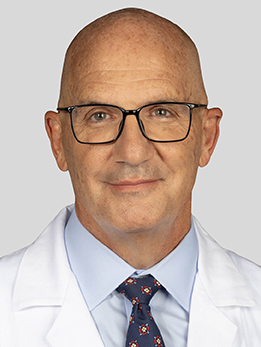
Dr. DiPatri has been performing pediatric neurosurgery for over 27 years. He's dual board-certified in both neurological surgery and pediatric neurological surgery, and he's Chief of Pediatric Neurosurgery for the Chicagoland Children's Health Alliance, meaning he oversees pediatric brain care across Comer, Advocate, and Endeavor Health. He came to UChicago after 22 years at Lurie Children's Hospital, with additional fellowship training at Boston Children's Hospital. Chiari malformation is one of the conditions Dr. DiPatri is most frequently consulted on — after 27 years of pediatric neurosurgery practice in Chicago, he has guided hundreds of families through the question of whether their child actually needs a decompression or just careful follow-up. He now leads the pediatric Chiari program at UChicago Medicine Comer Children's Hospital.

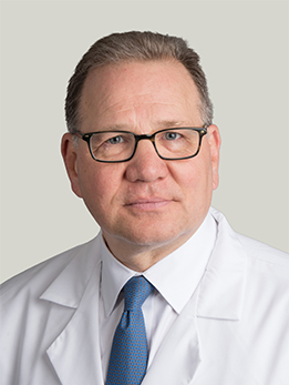
Dr. Das is Director of Neurotrauma at UChicago's Level 1 Trauma Center and directs the Neurosurgical Trauma Fellowship, one of only a few dedicated neurosurgical trauma fellowships in the country. She manages both head trauma and acute spine pathology and has been named to the Bucksbaum-Siegler Institute for Clinical Excellence. She trained at the University of Minnesota for residency and completed a skull base fellowship at Cleveland Clinic. Dr. Das performs posterior fossa decompression for children and adults with symptomatic Chiari I, and she is one of the surgeons at UChicago who will sit down with a family, review the imaging side by side, and walk through the bone-only versus duraplasty decision before recommending an operation.
Dr. Herman is Program Director of the Neurological Surgery Residency and a complex spine surgeon who practices the full spectrum of spine and neurorestoration procedures. He co-developed a fully implantable wireless intraspinal microstimulation device for restoring motor function after spinal cord injury, with publications in Artificial Organs and Scientific Reports. He has been named a Top Chicago Doctor for over a decade. Dr. Herman is one of the spine-focused neurosurgeons who sees Chiari patients at UChicago, particularly when a syrinx, scoliosis, or craniocervical instability is part of the picture and the case overlaps with complex pediatric and adult spinal care.
What Is a Chiari Malformation?
A Chiari malformation is a structural issue at the junction between the brain and the spinal cord. In a Chiari I, the lowest part of the cerebellum — two small lobes called the cerebellar tonsils — hangs more than 5 mm below the foramen magnum, the hole at the base of the skull where the spinal cord enters. In plain terms, there is a little less room than there should be at the back of the skull, and a piece of brain is sitting where it normally would not.
The consequence is mechanical. The crowded tonsils can block the normal back-and-forth flow of cerebrospinal fluid (CSF) between the head and the spine. When coughing, laughing, or straining briefly raises pressure inside the skull, that pressure cannot equalize smoothly — and the result is often the classic Chiari headache. Over time, blocked CSF flow can also push fluid into the center of the spinal cord and form a syrinx (also called syringomyelia), a thin cavity that can stretch and damage the cord from the inside.
The most important thing to know up front is that a Chiari I on an MRI is not the same thing as a Chiari problem. MRI scanners are sensitive enough that we now find mild tonsillar descent in children who have no symptoms at all. That distinction — between a finding on a scan and a condition that is actually causing harm — is the single most important judgment call in this diagnosis.
Although this article is focused on children, the same principles apply to adults. Chiari I is frequently diagnosed in the 20s, 30s, and 40s, sometimes after years of unexplained occipital headaches.
At a Glance
- A Chiari I malformation means the lower part of the cerebellum (the tonsils) sits more than 5 mm below the normal opening at the base of the skull
- Most children with a Chiari I found on MRI have no symptoms and never need surgery — watching is usually the right first step
- The classic symptom is a short, pounding headache at the back of the head that is triggered by coughing, laughing, or straining
- Surgery is recommended mainly for children with disabling Chiari-type headaches, a syrinx (fluid cyst) in the spinal cord, or neurologic problems from brainstem compression
- The best outcomes come from centers that operate on Chiari often, decide carefully who needs surgery, and follow children for years afterward
Have imaging or a diagnosis already?
We'll have a specialist review your MRI and records — often within 24 hours.
What Does It Feel Like?
Chiari I causes very specific patterns of symptoms — and many children with "tonsillar ectopia" on MRI have none of them. The details matter.
The classic Chiari headache
- A short, intense, pounding pain at the back of the head (the occipital region), sometimes spreading into the neck
- Triggered by coughing, laughing, sneezing, straining, heavy lifting, or bearing down — what neurologists call a tussive or Valsalva headache
- Lasts seconds to minutes and then fades
- Does not behave like a typical migraine or tension headache
Symptoms from a syrinx or brainstem pressure
- Numbness, tingling, or burning in the arms or hands
- Weakness or clumsiness in the hands, sometimes in a "cape" distribution over the shoulders
- A new curve of the spine (scoliosis), especially in younger children — sometimes the first sign of an underlying syrinx
- Neck pain
- Difficulty swallowing, a choking sensation, or a change in voice
- Sleep apnea, especially in infants and toddlers
- Balance problems, dizziness, or unsteady walking
What is usually NOT Chiari
Chronic daily headaches, migraines with aura, isolated dizziness, anxiety, or fatigue — even if an MRI shows a few millimeters of tonsillar descent — are rarely caused by Chiari. Operating on these symptoms does not reliably make them better, which is why we take a careful history before recommending surgery.
How Is It Diagnosed?
Diagnosis almost always starts with an MRI of the brain. On a sagittal (side view) image, the radiologist measures how far the cerebellar tonsils extend below a line drawn across the foramen magnum. More than 5 mm of descent in a child — with the right shape ("peg-like") and crowding at the craniocervical junction — is consistent with Chiari I.
If the brain MRI shows Chiari, the next step is almost always a full-spine MRI to look for a syrinx. This is important: a child can have a completely silent syrinx that is slowly stretching the spinal cord, and finding it changes the treatment plan significantly. We usually order the full spine MRI up front so we do not miss it.
A third, often underused test is a cine ("movie") MRI of CSF flow. This specialized sequence shows whether cerebrospinal fluid is actually moving freely across the foramen magnum or whether flow is obstructed. When symptoms are ambiguous, a cine MRI can help decide whether the Chiari is mechanically meaningful or just an incidental finding.
Other pieces of the workup may include:
- A sleep study in infants, toddlers, or any child with loud snoring, apneic spells, or daytime sleepiness — because brainstem compression can cause central sleep apnea
- Scoliosis films and a detailed neurologic exam
- A focused headache history to distinguish true Chiari-type headaches from migraine, tension, or medication-overuse headaches
At UChicago, we often re-read outside MRIs and add the spine images or cine flow study if they were not done. Getting the imaging right up front prevents unnecessary surgery — and catches the syrinxes that actually need it.
Types of Chiari Malformation
Despite the similar names, the different Chiari "types" are very different conditions. This article is about Chiari I, but it helps to know where it fits.
Chiari I
The most common form. The cerebellar tonsils sit more than 5 mm below the foramen magnum, but the brainstem and fourth ventricle are in a normal position. Usually diagnosed in childhood, adolescence, or early adulthood. Not associated with spina bifida. Most cases are managed conservatively; a minority need surgical decompression.
Chiari 1.5
An intermediate form first carefully described by Tubbs and colleagues. A Chiari 1.5 looks like a Chiari I on MRI (tonsils are herniated), but the brainstem is also pulled down through the foramen magnum. It matters because children with Chiari 1.5 are somewhat more likely to have persistent syringomyelia after a standard decompression and may need a more extensive operation or closer follow-up. Recognizing it on the preoperative scan changes the surgical plan.
Chiari II
A fundamentally different, congenital problem almost always seen in children born with myelomeningocele (open spina bifida). In Chiari II, not only the tonsils but also the brainstem, fourth ventricle, and part of the cerebellum are displaced down into the upper spinal canal, and the posterior fossa itself is abnormally small. Chiari II is usually diagnosed before or at birth, is managed as part of comprehensive spina bifida care, and is the target of fetal surgery: the MOMS trial showed that prenatal closure of a myelomeningocele reduces the need for shunting and partially reverses the hindbrain herniation compared to postnatal repair. Chiari II is its own condition with its own treatment pathway and is not the focus of this page.
And in adults
Although we specialize in the pediatric population here, Chiari I is common in adults too — and the principles described on this page (when to operate, bone-only versus duraplasty, syrinx management) apply across the age spectrum. Adult patients are seen by our adult neurosurgery team in the same health system.
How Is It Treated?
Step one: decide whether treatment is actually needed
The hardest and most important decision in Chiari care is who should have surgery in the first place. For children with a Chiari I found incidentally on an MRI — no headaches, no syrinx, a normal neurologic exam — the answer is usually no surgery, just watch. Large natural history studies of asymptomatic children show that new symptoms develop in only about 5–6% and a new syrinx in only 2–3% over years of follow-up. The international consensus for pediatric Chiari I endorses conservative management in this group. We typically see these children in clinic, make sure the spine MRI is clean, and plan a check-in rather than routine repeat imaging.
Surgery is generally recommended when a child has:
- Disabling, classic Chiari-pattern headaches (tussive / Valsalva-triggered occipital pain)
- A syrinx in the spinal cord, especially if it is growing or causing symptoms
- Neurologic signs from brainstem or cranial-nerve compression (swallowing difficulty, central sleep apnea, progressive weakness or sensory loss)
- Scoliosis linked to an underlying syrinx
Posterior fossa decompression
The operation is called a posterior fossa decompression. The child is positioned face-down, and through an incision at the back of the head and upper neck, the surgeon removes a small piece of the back of the skull (a suboccipital craniectomy) and usually the back arch of the first cervical vertebra (C1 laminectomy). That creates more room for the crowded cerebellum and restores normal CSF flow across the craniocervical junction.
At that point, the surgeon faces the central technical choice in Chiari surgery:
Bone-only decompression vs. duraplasty — an ongoing debate
Once the bone is removed, there are two main approaches:
- Bone-only (extradural) decompression. The tough outer lining of the brain (the dura) is left intact, or sometimes only its outer layer is scored. This is a shorter, less invasive operation with a faster recovery and fewer complications.
- Posterior fossa decompression with duraplasty (PFDD). The dura is opened and a patch (from the child's own tissue or a graft) is sewn in to expand the space further. This is a bigger operation with a higher rate of complications — CSF leak, pseudomeningocele, chemical meningitis — but it more reliably relieves symptoms and shrinks syrinxes.
Which approach is better is one of the most actively debated questions in pediatric neurosurgery, and there is no one-size-fits-all answer. The largest body of evidence on this question comes from the Park-Reeves Syringomyelia Research Consortium, a multicenter collaboration that has followed hundreds of pediatric Chiari patients prospectively. In their 692-patient study, bone-only decompression was associated with fewer complications and shorter hospital stays, but a higher reoperation rate and lower rates of headache and syrinx improvement compared to duraplasty. Duraplasty did more, at the cost of more complications — especially related to the dural opening itself.
The practical conclusion is that the right operation depends on the child in front of you: the size of the syrinx, the severity of symptoms, the anatomy on MRI, and the family's tolerance for reoperation versus complications. A center that performs both operations regularly — and talks through the trade-offs honestly — is the point of this conversation.
Syringo-subarachnoid shunts and revision surgery
A small number of children have a persistent or enlarging syrinx after a first operation. Options then include redoing the decompression with a more aggressive duraplasty, addressing arachnoid scarring directly, or placing a syringo-subarachnoid shunt — a small tube that drains the syrinx. These revision operations are uncommon and are best done at a center that sees enough Chiari to handle them.
Recovery
Most children stay in the hospital 2–4 nights after a Chiari decompression. Neck stiffness and a headache at the incision are expected for the first week or two. Most kids are back to school in 3–6 weeks and back to non-contact sports in a few months.
Considering surgery or planning a second opinion?
Our multidisciplinary team reviews complex cases together. You'll get a coordinated plan, not one opinion.
What Are the Outcomes?
For the right child, posterior fossa decompression works well. Large pediatric series report that around 80–90% of children who undergo decompression have meaningful improvement or resolution of their Chiari-type headaches, and published series find symptom resolution in roughly 90% of carefully selected patients. Syrinxes shrink in the large majority of children, with most of the reduction happening within the first 3 months after surgery and continuing to improve over the first year or two.
The picture is somewhat different for bone-only versus duraplasty operations. The table below summarizes the main trade-offs, drawn primarily from the Park-Reeves Syringomyelia Research Consortium and complementary single- and multicenter pediatric series.
| Outcome | Bone-only (PFD) | With duraplasty (PFDD) | What to know |
|---|---|---|---|
| Headache improvement | ~80% | ~90% | Duraplasty slightly more reliable for symptom relief |
| Syrinx shrinkage | Lower | Higher | Most regression happens within 3 months and continues for 1–2 years |
| Overall complications | Lower | Higher | Most dural complications are CSF leak, pseudomeningocele, aseptic meningitis |
| Hospital stay | Shorter | Longer | Typically 2 vs. 3–4 nights |
| Need for re-operation | Higher | Lower | More children who start with bone-only eventually need duraplasty |
The bigger picture: the single most important decision in Chiari care is not which operation, but whether to operate at all — and then picking the right technique for the right child. Both of those decisions depend on how many of these patients your team sees and how carefully they are followed over time. That is why choosing a center with a dedicated pediatric Chiari program matters as much as the surgery itself.
References
Have Questions About Chiari Malformation?
Our team is here to review your case, discuss your options, and help you take the next step.
Schedule: (773) 702-2123