Our Specialists for Clival Chordoma
Clival chordomas are rare enough that most neurosurgeons see only a handful in an entire career. At UChicago, our skull base team treats these tumors with a combined endoscopic endonasal and open approach, in close partnership with radiation oncologists who specialize in proton beam therapy.
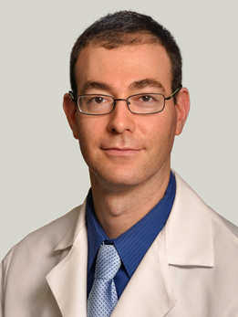
Dr. Horowitz is a skull base and neuro-oncology surgeon who also serves as Director of Quality and Associate Program Director for the Neurological Surgery residency. His laboratory research has identified novel genes driving meningioma and pediatric glioma formation, with work published in Nature Genetics and PNAS, and is funded by the DoD Neurofibromatosis Research Program. He holds a PhD in neuroscience from Northwestern and completed residency at Brigham and Women's/Boston Children's with a skull base fellowship at MD Anderson. Dr. Horowitz led the UChicago analysis of 736 skull base chordoma patients in the National Cancer Database, showing that endoscopic endonasal resection produces survival equivalent to open surgery while cutting hospital stay by roughly two days (Neurosurgical Focus, 2024). For most clival chordomas at UChicago, he is the surgeon planning and performing the endonasal resection.
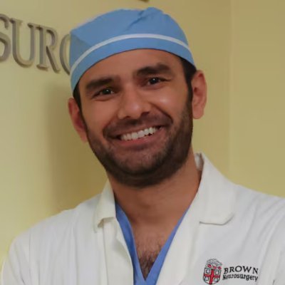
Dr. Ali is Director of Endoscopic Neurosurgery at UChicago, specializing in minimally invasive keyhole approaches to skull base and intracranial lesions. His practice focuses on endoscopic techniques for pituitary and skull base tumors, using narrow corridors through the nose or small cranial openings to reach deep lesions with minimal disruption of surrounding tissue. Dr. Ali works alongside Dr. Horowitz on complex endoscopic skull base cases, including clival chordomas that require combined endonasal and open approaches for lateral extension into the petrous bone or cavernous sinus.

Dr. Das is Director of Neurotrauma at UChicago's Level 1 Trauma Center and directs the Neurosurgical Trauma Fellowship, one of only a few dedicated neurosurgical trauma fellowships in the country. She manages both head trauma and acute spine pathology and has been named to the Bucksbaum-Siegler Institute for Clinical Excellence. She trained at the University of Minnesota for residency and completed a skull base fellowship at Cleveland Clinic. Dr. Das is first author on the largest modern epidemiologic study of chordoma in the United States, establishing the national incidence, demographics, and survival baseline that clinicians still cite when counseling newly diagnosed patients (Journal of Neuro-Oncology, 2020).
What Is a Clival Chordoma?
A clival chordoma is a rare, slow-growing bone tumor that arises in the clivus — the sloping wedge of bone at the base of the skull that sits directly in front of the brainstem. These tumors grow from tiny remnants of the notochord, a structure that guides spine development in the early embryo and then almost entirely disappears before birth. In a small number of people, a few notochord cells persist in the skull base or spine and, decades later, can give rise to a chordoma.
Chordomas are uncommon: about 1 case per million people per year in the United States. Roughly one-third of them arise in the skull base, and the clivus is the classic location. They can occur at any age but most often appear in adults in their 40s, 50s, and 60s, with a slight male predominance.
What makes clival chordomas so challenging is not how fast they grow — they are usually slow — but where they grow. The clivus sits in the middle of the most crowded real estate in the body: the brainstem behind it, the pituitary gland above it, the carotid arteries and cranial nerves on either side, and the upper airway below. A tumor that would be straightforward to remove almost anywhere else becomes a major operation when it is wrapped around these structures.
Chordomas are considered locally aggressive. They rarely spread to distant parts of the body early on, but they eat into bone, push on the brainstem, and come back again and again if any cells are left behind. That is why the goal of the first operation is so important, and why this is a tumor you want treated at a center that sees them regularly.
At a Glance
- Clival chordomas are rare tumors that grow from leftover notochord cells in the clivus, the bone at the base of the skull
- They are slow-growing but locally aggressive, pressing on the brainstem, cranial nerves, and major blood vessels
- The most important treatment is maximum safe surgical removal, usually through the nose using an endoscope
- Proton beam radiation is almost always given after surgery to kill any cells left behind
- They tend to come back even after good treatment, so lifelong MRI follow-up is essential
Have imaging or a diagnosis already?
We'll have a specialist review your MRI and records — often within 24 hours.
What Does It Feel Like?
Because the clivus is deep inside the head, early clival chordomas often cause no symptoms at all. Many are found when a patient has an MRI for an unrelated headache or ear problem. When symptoms do appear, they come from the tumor pressing on nearby nerves and structures.
Common early symptoms
- Headache, often dull and centered behind the eyes or at the back of the head
- Double vision (diplopia), usually from pressure on the sixth cranial nerve, which controls outward eye movement
- Neck pain or stiffness
- A muffled or full feeling in one ear
Symptoms as the tumor grows
- Difficulty swallowing or a hoarse voice
- Facial numbness or tingling
- Trouble with balance or walking
- Weakness on one side of the body
- Hormone changes if the tumor pushes on the pituitary gland
- In advanced cases, nasal obstruction or a mass visible in the back of the throat
Double vision from a new sixth-nerve palsy in an adult is one of the most common reasons a clival chordoma is eventually found. If you have developed sudden or unexplained double vision, ask your doctor whether an MRI of the brain and skull base is appropriate.
How Is It Diagnosed?
The workup for a suspected clival chordoma almost always starts with imaging and ends with tissue.
MRI with contrast
An MRI of the brain and skull base with gadolinium contrast is the most important test. Chordomas have a characteristic appearance: they are typically bright on T2-weighted images, destroy bone, and often push the brainstem backward. MRI also shows how close the tumor is to the carotid arteries, cranial nerves, and pituitary gland — information your surgical team needs to plan the safest approach.
CT scan
A high-resolution CT scan complements the MRI by showing exactly how much of the clivus and surrounding bone the tumor has eaten through. CT is also used to build the navigation plan that guides the surgeon during the operation.
Biopsy and pathology
The only way to confirm a chordoma is to examine tumor tissue under a microscope. In most cases, the biopsy and the main surgery are done in the same operation. Pathologists look for the classic physaliphorous cells — plump cells with a bubbly-looking cytoplasm that sit in a gel-like matrix.
Brachyury testing
The single most important test for confirming chordoma is staining for brachyury, a transcription factor that is expressed in notochord cells and in essentially every conventional chordoma. In large series, nuclear brachyury staining is positive in more than 95% of chordomas and is almost never seen in the tumors that look like chordoma under the microscope, such as chondrosarcoma. Brachyury is what turns a suggestive MRI and ambiguous pathology into a confident diagnosis.
The difference that matters: chordoma vs. chondrosarcoma
Clival chondrosarcomas can look almost identical to chordomas on imaging but behave very differently — they respond better to radiation and have markedly better long-term survival. Distinguishing the two is one of the most important jobs of the pathology team, and brachyury is the key.
Types of Clival Chordoma
Under the 2020 World Health Organization classification of bone tumors, clival chordomas are divided into three histologic subtypes. They can look similar on imaging but behave very differently.
Conventional chordoma
By far the most common type — roughly 80-90% of clival chordomas. Made up of cords and lobules of classic physaliphorous cells in a myxoid (mucus-like) matrix. Strongly positive for brachyury. Slow-growing but prone to local recurrence.
Chondroid chordoma
A variant in which parts of the tumor look like cartilage. Historically thought to have a better prognosis than conventional chordoma, though modern series suggest the difference is smaller than once believed. Still brachyury-positive. Must be carefully distinguished from chondrosarcoma, which has a genuinely better outlook.
Dedifferentiated chordoma
The rarest and most aggressive subtype — about 2-5% of cases. Here a conventional chordoma is juxtaposed with a high-grade sarcoma that has lost brachyury expression and often carries TP53 mutations. Dedifferentiated chordomas can metastasize, grow rapidly, and historically have a median overall survival of about 20 months. They are usually treated with a combination of aggressive surgery, radiation, and sometimes systemic therapy in clinical trials.
A newer category, poorly differentiated chordoma, has been recognized over the last decade. It affects mostly children and young adults, is defined by loss of the SMARCB1/INI-1 gene, and behaves more aggressively than conventional chordoma. It is uncommon in the clivus of older adults but important to recognize in younger patients.
How Is It Treated?
Treatment of a clival chordoma is a carefully sequenced combination of maximum safe surgery followed by high-dose radiation, usually with proton beams. Chemotherapy has historically played a limited role, though brachyury-targeted therapies are an active area of research.
Surgery: the endoscopic endonasal approach
For most clival chordomas, the endoscopic endonasal approach — operating through the nostrils with a long, thin camera and specialized instruments — has become the workhorse operation. Instead of opening the skull, the surgeon reaches the clivus directly through the back of the nose and the sphenoid sinus. This gives a head-on view of the tumor, preserves the brainstem and cranial nerves by working in front of them rather than around them, and avoids any external incision or brain retraction.
A UChicago-led analysis of 736 patients in the National Cancer Database showed that endoscopic endonasal resection is now used in about 62% of skull base chordoma operations in the United States, is used more often every year, and achieves survival outcomes equivalent to open surgery while shortening hospital stay by about two days.
Expanded and open approaches
When a tumor extends far to the side — into the petrous bone, the cavernous sinus, or around the vertebral arteries — an endoscopic approach alone may not reach all of it safely. In those cases, the team may use an expanded endonasal approach, combine the endoscopic route with a far-lateral or transcondylar open approach, or plan a staged operation that tackles different parts of the tumor in separate sittings. The choice depends entirely on the shape of the tumor and which structures are in the way.
Reconstruction and CSF leak prevention
A skull base opening into the nose has to be watertight to keep cerebrospinal fluid (CSF) out of the sinuses. Your surgical team uses a nasoseptal flap — a piece of well-vascularized lining from the inside of the nose — to cover the defect, often combined with fat, fascia, and a lumbar drain. Modern reconstruction has brought CSF leak rates after endoscopic clival surgery down to roughly 5-10% in experienced hands.
Proton beam radiation therapy
After surgery, nearly every patient with a clival chordoma receives high-dose proton beam radiation therapy. Chordomas are relatively resistant to radiation and need very high doses (typically 70-79 Gy) for durable control — doses that are hard to deliver with conventional photon radiation without damaging the brainstem, optic nerves, and temporal lobes. Protons can be aimed more precisely and stop at a defined depth, concentrating the dose on the tumor bed while sparing normal tissue.
In published series, proton therapy after surgery produces 5-year local control rates of 70-80% and 5-year overall survival rates of roughly 80%, substantially better than surgery alone. Proton therapy is now the standard of care after clival chordoma resection at essentially every major skull base center.
Chemotherapy and targeted therapy
Traditional chemotherapy does not work well against chordoma. For tumors that recur or cannot be fully treated, current options include targeted drugs such as imatinib (which blocks PDGFR) and, in clinical trials, agents targeting brachyury, the EGFR pathway, and tumor immunology. A randomized phase II trial of the yeast-brachyury vaccine GI-6301 in combination with radiation did not show a clear survival benefit, but brachyury remains one of the most promising drug targets in chordoma and multiple next-generation approaches are in development.
When the tumor comes back
Recurrence is common, especially in tumors that could not be completely removed the first time. The usual approach is another operation to remove as much recurrent disease as possible, followed by additional radiation if safe, or enrollment in a clinical trial. Every recurrence conversation should involve a multidisciplinary team that includes neurosurgery, radiation oncology, medical oncology, and pathology.
Considering surgery or planning a second opinion?
Our multidisciplinary team reviews complex cases together. You'll get a coordinated plan, not one opinion.
What Are the Outcomes?
Outcomes for clival chordoma depend on three things above all: how much of the tumor is removed at the first operation, whether the patient receives adjuvant proton beam radiation, and the histologic subtype. The best results come from a complete or near-complete resection followed by proton therapy at a high-volume skull base center.
| Scenario | 5-Year Overall Survival | 5-Year Progression-Free | What to know |
|---|---|---|---|
| Gross total resection + proton therapy | ~80-90% | ~65-75% | The best achievable outcome |
| Subtotal resection + proton therapy | ~65-75% | ~40-55% | Most common real-world scenario |
| Surgery alone, no adjuvant radiation | ~50% | ~25-35% | Much higher recurrence rate |
| Conventional / chondroid subtype | ~75-85% | ~55-70% | Typical clinical outcome |
| Dedifferentiated chordoma | ~20-30% | Often <12 months | Median OS historically ~20 months |
How chordomas recur
Unlike many other cancers, chordoma recurrences are almost always local — they show up in or next to the original tumor bed rather than spreading to other parts of the body. Distant metastases (to the lungs, liver, or spine) happen in fewer than 10-20% of patients, and almost always late in the disease. This is why repeat MRIs every 6-12 months for the rest of your life are a core part of follow-up care, and why catching a small recurrence early can make a second surgical cure possible.
Why experience matters
Published series consistently show that patients treated at high-volume skull base centers have higher rates of complete resection, lower complication rates, and better long-term survival than those treated at low-volume centers. Clival chordomas are rare enough that no single surgeon sees very many of them, which is why the best outcomes come from a team that has standardized how it approaches this tumor — from the first MRI, through reconstruction, to proton therapy, to decades of follow-up.
References
Have Questions About Clival Chordoma?
Our team is here to review your case, discuss your options, and help you take the next step.
Schedule: (773) 702-2123