Our Specialists for Craniopharyngioma
Craniopharyngioma surgery is one of the clearest examples of a condition where experience and subspecialty expertise change outcomes. At UChicago, these cases are managed by a dedicated skull base and pituitary team who decide between endoscopic and transcranial approaches based on the individual anatomy of the tumor, not the comfort zone of the surgeon.
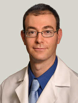
Dr. Horowitz is a skull base and neuro-oncology surgeon who also serves as Director of Quality and Associate Program Director for the Neurological Surgery residency. His laboratory research has identified novel genes driving meningioma and pediatric glioma formation, with work published in Nature Genetics and PNAS, and is funded by the DoD Neurofibromatosis Research Program. He holds a PhD in neuroscience from Northwestern and completed residency at Brigham and Women's/Boston Children's with a skull base fellowship at MD Anderson. Dr. Horowitz co-directs the UChicago Pituitary and Neuroendocrine Disorders Program and completed a skull base fellowship at MD Anderson specifically focused on tumors like craniopharyngioma. He is one of the surgeons patients are most likely to see for a sellar or suprasellar mass at UChicago, and his lab has published on the genetic drivers of related brain tumors in Nature Genetics and PNAS.
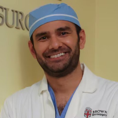
Dr. Ali is Director of Endoscopic Neurosurgery at UChicago, specializing in minimally invasive keyhole approaches to skull base and intracranial lesions. His practice focuses on endoscopic techniques for pituitary and skull base tumors, using narrow corridors through the nose or small cranial openings to reach deep lesions with minimal disruption of surrounding tissue. Dr. Ali is Director of Endoscopic Neurosurgery at UChicago and specializes in the endoscopic endonasal approach to sellar and suprasellar tumors. For a craniopharyngioma with favorable midline anatomy, he is the surgeon who operates through the nose — giving a direct, below-the-chiasm view of the tumor with no scalp incision.
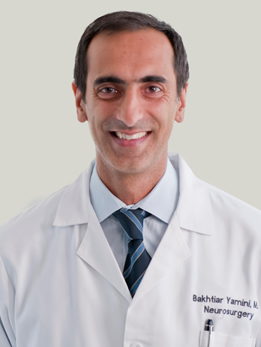
Dr. Yamini is a brain tumor surgeon and scientist who serves as Vice Chair for Academic Affairs and Director of Neurosurgical Oncology at UChicago. In the operating room, he uses advanced imaging and navigation tools for stereotactic biopsy, laser ablation, and image-guided maximal resection. In his lab, he runs NIH-funded research into why some tumors resist treatment and into biodegradable nanoparticle vectors that deliver drugs directly to CNS tumors. Dr. Yamini is Director of Neurosurgical Oncology and Vice Chair for Academic Affairs, and he brings the tumor-biology side of craniopharyngioma care into the operating room: his NIH-funded lab studies treatment resistance in CNS tumors and biodegradable nanoparticle drug delivery, and he is one of the surgeons who integrates molecular testing (BRAF, CTNNB1) into decisions about adjuvant therapy.
What Is a Craniopharyngioma?
A craniopharyngioma is a slow-growing tumor that forms in the sellar and suprasellar region — the area just above and behind your nose where the pituitary gland sits, right beneath the optic nerves and hypothalamus. They arise from leftover cells of a structure called Rathke's pouch, which in the embryo helps form the pituitary gland.
Under a microscope, craniopharyngiomas are benign. They don't invade distant tissues and they don't spread through the body. But doctors often describe their behavior as aggressive, and for good reason: they grow in the most crowded neighborhood in the brain. Even a small tumor can push on the optic nerves, strangle the pituitary stalk, and damage the hypothalamus — the small but critical region that regulates hunger, thirst, temperature, sleep, and the stress response.
Craniopharyngiomas are uncommon. They account for roughly 1–3% of all brain tumors, with an incidence of about 0.16 per 100,000 people per year in the United States. They show up at any age but have a distinctive bimodal pattern: one peak in children aged 5–14, and a second peak in adults aged 50–74.
At a Glance
- Craniopharyngiomas are benign tumors but behave aggressively because of where they sit — next to the optic nerves, pituitary, and hypothalamus
- They occur in two age peaks: children aged 5–14 and adults aged 50–74
- The two main types — adamantinomatous and papillary — are now known to be genetically distinct diseases with different treatment implications
- Surgery is the primary treatment, with the choice between endoscopic endonasal and transcranial approaches driven by tumor anatomy
- Papillary craniopharyngiomas carry a BRAF V600E mutation in about 95% of cases, and a targeted drug combination can shrink them without surgery in many patients
Have imaging or a diagnosis already?
We'll have a specialist review your MRI and records — often within 24 hours.
What Does It Feel Like?
Because craniopharyngiomas grow where three critical systems converge — vision, hormones, and the fluid spaces of the brain — the symptoms usually fall into three buckets. Most patients have at least two of them by the time they're diagnosed.
Vision changes
The tumor sits directly under the optic chiasm, so pressure on the nerves there is one of the most common presentations.
- Gradual loss of peripheral vision, especially to the outer sides (bitemporal hemianopsia — a pattern that feels like wearing blinders)
- Blurred or dim vision in one or both eyes
- New trouble with depth perception or bumping into things on the side
- In children, a lazy eye or a child who stops noticing objects off to the side
Hormone problems
The tumor compresses the pituitary stalk and gland, which disrupts the body's hormonal signaling.
- Fatigue, low energy, and cold intolerance (from low thyroid or cortisol)
- Excessive thirst and urination, sometimes many liters a day (diabetes insipidus)
- Stalled growth or delayed puberty in children
- Loss of menstrual periods, erectile dysfunction, or low libido in adults
- Unexplained weight gain, especially when the hypothalamus is involved
Pressure and hypothalamic symptoms
Larger tumors can block the flow of cerebrospinal fluid and cause pressure to build inside the skull.
- Morning headaches, sometimes with nausea or vomiting
- Confusion, memory changes, or personality shifts
- Disrupted sleep, temperature regulation, or appetite control
- In infants and young children, an abnormally growing head, irritability, or missed developmental milestones
How Is It Diagnosed?
The workup for a suspected craniopharyngioma moves quickly once symptoms point toward the pituitary region. The goal is to confirm the diagnosis, map the tumor's exact relationship to nearby structures, and measure how much damage has already been done to hormones and vision — all before surgery.
MRI with contrast
The single most important test is a dedicated pituitary MRI with contrast. Craniopharyngiomas have a fairly distinctive appearance: a mix of solid tissue and fluid-filled cysts, often with calcium deposits. On MRI, the cysts may look bright on certain sequences because of the oily, cholesterol-rich fluid inside them. A high-quality MRI also tells your surgical team exactly how the tumor relates to the optic nerves, carotid arteries, pituitary stalk, and hypothalamus — information that drives the choice of surgical approach.
CT scan
A CT scan is usually added to confirm calcifications, which are present in most adamantinomatous craniopharyngiomas and help distinguish them from other sellar tumors like pituitary adenomas or Rathke's cleft cysts.
Hormone testing and endocrine workup
Before any surgery, you'll have a full panel of pituitary hormone tests. This includes cortisol, thyroid, growth hormone, prolactin, sex hormones, and measures of water balance. Many patients turn out to have deficiencies they didn't know about, and any missing hormones — especially cortisol — need to be replaced before the operating room.
Formal visual field testing
An ophthalmologist will document your baseline vision with acuity measurements, a dilated eye exam, and automated visual field testing. This gives the surgical team a precise map of what was lost before surgery and a baseline to measure recovery against.
Pathology and molecular testing
The definitive diagnosis comes after surgery, when a neuropathologist examines the tissue. The pathologist will also test for the tumor's molecular signature — specifically, whether it carries a BRAF V600E mutation (pointing to the papillary subtype) or a CTNNB1 (beta-catenin) mutation (pointing to the adamantinomatous subtype). These findings are no longer academic: they guide decisions about radiation and targeted drug therapy.
Types of Craniopharyngioma
For decades, craniopharyngiomas were lumped together as one tumor. We now know they are two biologically distinct diseases that happen to grow in the same spot. The distinction matters because it changes how we think about surgery, radiation, and new drug options.
Adamantinomatous craniopharyngioma
This is the classic subtype and by far the most common in children, though it also occurs in adults. Under the microscope, it looks like a tooth bud — irregular sheets of cells, wet keratin, and cholesterol-filled cysts that give it the nickname "machine oil" fluid. These tumors almost always carry a mutation in the CTNNB1 gene, which encodes beta-catenin, driving a hyperactive Wnt signaling pathway.
- Most common in children aged 5–14, but seen at all ages
- More likely to be heavily calcified and cystic
- Tends to stick more aggressively to the hypothalamus and surrounding brain, which raises the risk of injury during complete removal
- Recurrence rates are higher, especially after incomplete removal
Papillary craniopharyngioma
This subtype is almost exclusively seen in adults, usually in the 5th–7th decades of life. It looks very different under the microscope — firm, well-circumscribed, and usually solid rather than cystic. About 95% of papillary craniopharyngiomas carry a BRAF V600E mutation, the same mutation that drives melanoma and some thyroid cancers.
- Almost exclusively adult onset
- Usually solid, less calcified, and often better demarcated from surrounding brain
- The BRAF V600E mutation makes them responsive to BRAF/MEK inhibitor drugs — an option that in some patients can shrink the tumor dramatically without a scalpel
Knowing the subtype before or early in treatment can open doors. An adult with a BRAF-mutant papillary craniopharyngioma in a hostile anatomic location may be a candidate for drug therapy first, shrinking the tumor before any surgery is attempted.
How Is It Treated?
Craniopharyngioma treatment is a balancing act. On one side is the tumor — the surgeon wants to remove as much as possible, because residual tumor is the single biggest driver of recurrence. On the other side are the optic nerves, pituitary stalk, and hypothalamus, which can tolerate only so much handling before vision, hormones, and quality of life pay the price. Modern care has moved decisively toward protecting function first and using radiation or targeted drugs to mop up anything left behind.
Surgery: endoscopic endonasal vs. transcranial
Surgery is the first and most important step for almost every patient. There are two main surgical corridors, and the right choice depends on the tumor's anatomy — not the surgeon's preference.
Endoscopic endonasal approach (EEA). The surgeon reaches the tumor through the nose, with no scalp incision, using a high-definition endoscope. For tumors that sit midline, in or just above the sella, this approach gives a direct view from below the optic nerves — which means the surgeon isn't working around the nerves to reach the tumor. Published comparative studies show that when both approaches are feasible, endoscopic endonasal surgery is associated with higher rates of gross total resection, better visual recovery, and less visual deterioration than transcranial surgery, at the cost of a higher risk of cerebrospinal fluid leak — a complication that has been dramatically reduced with modern skull base closure techniques.
Transcranial (craniotomy) approach. For tumors that extend far laterally, wrap around major arteries, or push high up into the third ventricle, a traditional craniotomy may still be the safer choice. Transcranial surgery gives a wider panoramic view of the suprasellar region and better access to tumors far from the midline.
At UChicago, the decision between approaches is made collaboratively by surgeons who are comfortable with both. That matters: in many centers, the approach is chosen by default based on which surgeon is available, not which approach fits the tumor.
The trade-off that defines craniopharyngioma surgery
Decades of published data have shown that aggressive attempts to remove every last bit of tumor — when it's fused to the hypothalamus — can cause devastating hypothalamic injury. Patients (especially children) can develop uncontrolled weight gain, personality change, cognitive slowing, and disrupted temperature and sleep regulation. For this reason, the modern philosophy is hypothalamus-sparing surgery: remove as much as safely possible, leave what's stuck to the hypothalamus, and use radiation afterward to control the small amount that remains.
Adjuvant radiation for residual or recurrent tumor
When a small amount of tumor is intentionally or unavoidably left behind, focused radiation is highly effective at controlling it. Options include:
- Fractionated photon radiation therapy (standard external beam), delivered over several weeks
- Proton beam therapy, which spares surrounding brain tissue and is especially valuable in children. A recent prospective phase 2 study (RT2CR, Lancet Oncology 2023) showed excellent tumor control with limited surgery plus proton therapy in children and adolescents
- Stereotactic radiosurgery for small, well-defined recurrences at a safe distance from the optic nerves
Studies consistently show that adjuvant radiation after subtotal resection produces local control rates comparable to gross total resection, but with far less hypothalamic injury — the reason it has become the standard of care.
BRAF-targeted therapy for papillary craniopharyngioma
This is one of the most important recent advances in neuro-oncology. In a landmark 2023 New England Journal of Medicine trial, 15 of 16 patients with newly diagnosed, BRAF-mutant papillary craniopharyngiomas had a partial response or better to the combination drug regimen vemurafenib plus cobimetinib, with many tumors shrinking by more than 90%. For carefully selected adult patients with BRAF-mutant tumors, drug therapy can be considered before surgery or radiation — a paradigm shift for a disease that used to be managed with a scalpel alone.
Recurrence
Even with modern surgery and radiation, craniopharyngiomas can come back. When they do, the treatment options depend on where the recurrence sits: repeat surgery, additional radiation, cyst drainage with an Ommaya reservoir, or — for BRAF-mutant papillary tumors — targeted drug therapy. Lifelong surveillance with MRI and endocrine testing is essential.
Considering surgery or planning a second opinion?
Our multidisciplinary team reviews complex cases together. You'll get a coordinated plan, not one opinion.
What Are the Outcomes?
Long-term survival for craniopharyngioma is generally high — overall survival at 10 years ranges from 80% to over 95% depending on age at diagnosis, extent of resection, and hypothalamic involvement. But survival is only one part of the story. For this tumor more than almost any other, the conversation has to include vision, hormones, and day-to-day quality of life.
| Outcome | Typical result | What drives it |
|---|---|---|
| 10-year overall survival | ~80–95% | Age, hypothalamic involvement, center experience |
| Recurrence after gross total resection | ~10–20% | Adherence to hypothalamus, subtype |
| Recurrence after subtotal resection alone | ~60–75% | Adjuvant radiation dramatically reduces this |
| Recurrence after subtotal resection + radiation | ~10–20% | Comparable to complete resection |
| Visual improvement after surgery | ~50–70% (higher with endoscopic) | Preoperative severity, approach, surgeon experience |
| New permanent pituitary hormone loss | >80% need lifelong replacement | Nearly universal after aggressive resection |
| Hypothalamic obesity | ~30–50% with hypothalamic injury | Main driver of long-term quality of life |
Quality of life — the part that really matters
For patients who survive craniopharyngioma, the biggest long-term challenge is usually not the tumor but the hypothalamic syndrome — a constellation of weight gain, disrupted sleep, fatigue, and impaired temperature and appetite regulation that can follow injury to the hypothalamus. Research from large European cohorts has shown that 20-year survival is lower in patients with hypothalamic involvement than in those without, and that quality of life is strongly determined by how much hypothalamic function is preserved.
This is why the choice of surgical team matters so much. The difference between a surgeon who knows when to stop at the hypothalamus — and uses radiation or targeted therapy to finish the job — and one who pushes for complete removal at all costs can change what the next 30 years of a patient's life look like.
References
Have Questions About Craniopharyngioma?
Our team is here to review your case, discuss your options, and help you take the next step.
Schedule: (773) 702-2123