Our Specialists for Glioblastoma
Glioblastoma is a race against a fast-moving tumor, and outcomes hinge on how much tumor can be safely removed in the first operation. The UChicago team handles glioblastoma every week, combines awake mapping with intraoperative imaging, and runs investigator-initiated clinical trials that are not available at most hospitals.
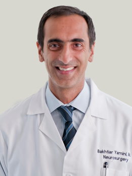
Dr. Yamini is a brain tumor surgeon and scientist who serves as Vice Chair for Academic Affairs and Director of Neurosurgical Oncology at UChicago. In the operating room, he uses advanced imaging and navigation tools for stereotactic biopsy, laser ablation, and image-guided maximal resection. In his lab, he runs NIH-funded research into why some tumors resist treatment and into biodegradable nanoparticle vectors that deliver drugs directly to CNS tumors. Dr. Yamini's laboratory at UChicago studies how NF-kB signaling drives temozolomide resistance in glioblastoma, and he is the principal investigator of a multi-institutional phase I trial adding acetazolamide to temozolomide for newly diagnosed MGMT-methylated patients (Neuro-Oncology Advances, 2024). If you want a surgeon who is also actively running glioblastoma clinical trials, he is the one to meet.
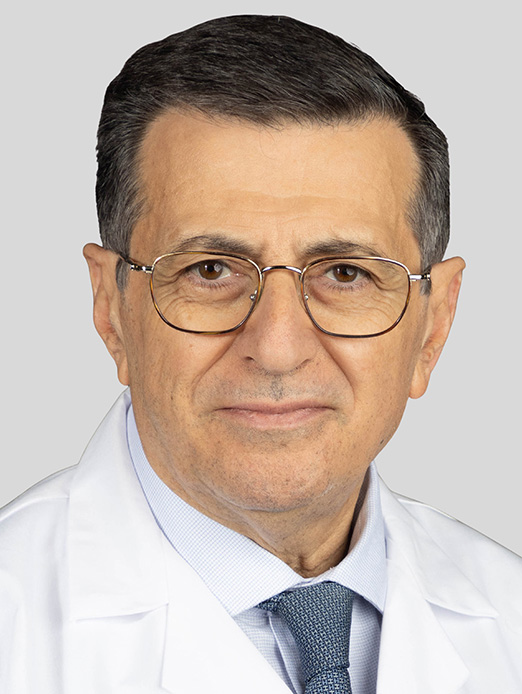
Dr. Comair is Section Chief of Neurosurgical Oncology and a pioneer of awake craniotomy for tumors in eloquent cortex. He has authored over 120 peer-reviewed publications and two neurosurgery textbooks, and he founded the first comprehensive epilepsy surgery program in the Middle East and North Africa. He trained at the Montreal Neurological Institute and previously held faculty positions at Cleveland Clinic, UCLA, Johns Hopkins, Baylor, and the American University of Beirut. For glioblastomas that present with seizures or sit near language or motor cortex, Dr. Comair brings decades of experience in awake craniotomy and intraoperative brain mapping — a skill set he developed leading complex tumor and epilepsy surgery programs before joining UChicago. He is one of the surgeons patients meet when a tumor looks "inoperable" somewhere else.
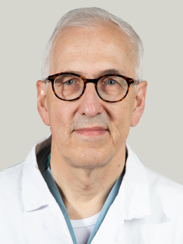
Dr. Warnke is an international leader in functional neurosurgery and has performed over 6,000 stereotactic surgeries and more than 3,000 brain tumor surgeries. He is only the second neurosurgeon worldwide to perform laser hemispherotomy, and he has completed over 400 laser ablation surgeries since arriving at UChicago. He is funded by four NIH grants including the BRAIN Initiative, and he directs the NAUTILUS trial for thalamic stimulation in drug-resistant epilepsy. Dr. Warnke's early work helped define when stereotactic biopsy plus radiation was the right approach for deep glioblastomas that could not be safely resected (J Neurosurg, 1993). At UChicago he now directs stereotactic and functional neurosurgery and uses laser interstitial thermal therapy (LITT) for small or deep recurrent glioblastomas that would otherwise require a second open craniotomy.
What Is Glioblastoma?
Glioblastoma (often called GBM) is a fast-growing cancer that starts in the supportive cells of the brain called glial cells. Under the 2021 World Health Organization classification, glioblastoma is defined as an IDH-wildtype, WHO grade 4 astrocytoma in adults. In plain language: it is the most aggressive form of primary brain tumor, and it behaves differently from the slower-growing gliomas that share part of its name.
About 12,000 Americans are diagnosed with glioblastoma every year. It is the most common malignant brain tumor in adults, making up roughly half of all primary malignant brain tumors. It can happen at any age, but it is most common in adults between 55 and 75.
Glioblastoma is aggressive for two reasons. First, the tumor cells divide quickly and build their own blood supply, which is why the tumor is often surrounded by swelling on MRI. Second, the tumor sends microscopic fingers of cancer cells out into the normal-looking brain around it, which is why even a so-called complete resection is almost never truly complete on a cellular level. This is also why surgery alone is not enough and why radiation and chemotherapy are always part of the plan.
Here is the part that matters most: glioblastoma is serious, but it is also treatable. Median survival with modern care is longer than it used to be, and at high-volume centers with experienced surgeons and clinical trial access, a meaningful minority of patients live years, not months.
At a Glance
- Glioblastoma is the most common aggressive primary brain tumor in adults, with about 12,000 new cases a year in the U.S.
- The standard first treatment is surgery to remove as much tumor as safely possible, followed by six weeks of radiation with daily temozolomide chemotherapy.
- How much of the tumor is removed in the first surgery is one of the single biggest factors in how long patients live.
- A genetic marker called MGMT methylation predicts who will respond best to temozolomide and roughly doubles median survival when present.
- Patients treated at high-volume academic centers with clinical trial access live meaningfully longer than those treated elsewhere.
Have imaging or a diagnosis already?
We'll have a specialist review your MRI and records — often within 24 hours.
What Does It Feel Like?
Glioblastoma symptoms come on over weeks, not years. If you or a family member had a sudden change in speech, strength, or thinking that led to an MRI, you are not imagining how fast this happened. The symptoms depend on where in the brain the tumor sits.
The most common first symptoms
- New or worsening headaches, often worse in the morning or when lying flat
- A seizure — sometimes a full convulsion, sometimes just a strange smell, a feeling of déjà vu, or a few seconds of staring
- Weakness, numbness, or clumsiness on one side of the body
- Trouble finding words, slurred speech, or difficulty understanding what people are saying
- Changes in personality, memory, or judgment that family members notice first
- Vision changes, especially a loss of peripheral vision on one side
- Nausea and vomiting without another explanation, caused by pressure inside the skull
Many of these symptoms can be caused by things that are not cancer. But when two or more of them appear together over a few weeks, a brain MRI is the right next step.
How Is It Diagnosed?
The path to a glioblastoma diagnosis usually happens fast, often within a few days of the first symptom.
Step 1: MRI with contrast
An MRI with gadolinium contrast is the test that first raises the suspicion. Glioblastoma has a characteristic look — an irregular, ring-enhancing mass with a dark center (that dark center is dead tumor tissue, called necrosis) surrounded by swelling. Your team will also look at advanced sequences like perfusion imaging and MR spectroscopy, which can help distinguish glioblastoma from other conditions like an abscess or a single brain metastasis.
Step 2: Surgery and tissue diagnosis
An MRI can strongly suggest glioblastoma, but the diagnosis is only made once a pathologist looks at actual tumor tissue under a microscope. In almost all cases, your neurosurgeon will recommend surgery as both the diagnosis and the first treatment in the same step — removing as much tumor as possible and sending it to pathology. In rare cases where the tumor sits in a location too risky to resect (deep in the brainstem, for example), a stereotactic needle biopsy is done instead to get tissue.
Step 3: Molecular and genetic testing
This is where modern glioblastoma care looks very different from what it did ten years ago. After the tumor is removed, the tissue is tested for specific molecular markers that affect both your prognosis and which treatments will work best:
- IDH status — glioblastoma is, by definition, IDH-wildtype. If the tumor is IDH-mutant, it is now classified as a different (and generally less aggressive) tumor called astrocytoma grade 4.
- MGMT promoter methylation — this is the single most important predictor of how well temozolomide will work. Patients with MGMT-methylated tumors live significantly longer on the Stupp protocol than patients without methylation.
- TERT promoter mutation, EGFR amplification, and chromosome 7 gain / 10 loss — these are additional features that confirm the diagnosis and, in research settings, can guide clinical trial eligibility.
You should make sure your tumor was sent for full molecular testing. If you're getting a second opinion, this is one of the first things an experienced team will ask about.
Types of Glioblastoma
Since the 2021 WHO update, all adult glioblastomas are, by definition, IDH-wildtype. In older textbooks you may have read about primary versus secondary glioblastoma, or about IDH-mutant glioblastoma. Those categories have been retired — what used to be called secondary or IDH-mutant glioblastoma is now classified as astrocytoma, IDH-mutant, WHO grade 4, which has a meaningfully better prognosis and is treated on a different pathway.
Within adult IDH-wildtype glioblastoma, the most important split is based on MGMT promoter methylation status, because it changes both prognosis and treatment planning.
MGMT methylated
About 40% of glioblastomas have a silenced (methylated) MGMT gene. MGMT is a DNA repair enzyme — when it is silenced, the tumor cannot repair the damage caused by temozolomide, which means the chemotherapy works much better. Patients with MGMT-methylated tumors have roughly double the median survival of patients with unmethylated tumors when treated with the Stupp protocol, and they are also the group most likely to benefit from Tumor Treating Fields.
MGMT unmethylated
The remaining 60% of patients have an active (unmethylated) MGMT gene and get less benefit from temozolomide. Standard of care is still surgery, radiation, and temozolomide, but this is also the group for whom clinical trials should be discussed up front, because the marginal gain from temozolomide alone is smaller.
Knowing which group you are in does not change whether you should treat your tumor aggressively — it changes how aggressively your team should be pushing you toward trials, how they dose your chemotherapy, and what they tell you to expect.
How Is It Treated?
Step 1: Maximal safe surgical resection
Surgery is the first and most important step. The goal is not just to get tissue for a diagnosis — it is to remove as much tumor as can be safely removed without damaging the parts of the brain that control movement, speech, vision, and personality. Landmark data from UCSF showed that every additional 5-10% of tumor removed translates into a measurable survival benefit, with the biggest jumps once you cross 78% and 95% resection thresholds (Journal of Neurosurgery, 2011). In plain language: who does your surgery matters.
At UChicago, several tools are used to push resection further without injuring healthy brain:
- Awake craniotomy with brain mapping — if the tumor sits near areas that control speech or movement, the patient is woken up during part of the surgery and asked to talk, name objects, or move a hand while the surgeon stimulates the cortex and maps out which areas must be protected. This allows resection right up to the edge of critical brain, not a centimeter away from it.
- 5-ALA fluorescence (pink drink) — a dye that makes glioblastoma cells glow pink under a special microscope light, letting the surgeon see tumor that would otherwise look like normal brain (Lancet Oncology, 2006).
- Intraoperative MRI and neuronavigation — real-time imaging that shows the surgeon how much tumor is left before closing.
- Laser interstitial thermal therapy (LITT) — for deep tumors that cannot be safely opened up, a thin laser probe is placed through a tiny hole in the skull and uses heat to destroy the tumor under MRI guidance.
Step 2: The Stupp protocol (radiation plus temozolomide)
After surgery, the established standard of care is the Stupp protocol, named after the oncologist who led the pivotal trial in 2005. It has two phases:
- Concurrent phase (about 6 weeks): focused radiation therapy to the tumor bed, Monday through Friday, along with a low daily dose of oral temozolomide chemotherapy (including weekends).
- Adjuvant phase (6 months): a rest for a few weeks, then six cycles of higher-dose temozolomide taken for five days out of every 28.
Stupp and colleagues showed in the New England Journal of Medicine that adding temozolomide to radiation increased median survival from 12.1 to 14.6 months and doubled two-year survival (NEJM, 2005). Five-year follow-up confirmed a lasting benefit, especially in patients with MGMT-methylated tumors.
Step 3: Tumor Treating Fields (Optune)
Tumor Treating Fields (TTFields), sold under the brand name Optune, is a wearable device that delivers low-intensity alternating electric fields through transducer arrays worn on a shaved scalp. It sounds strange, but in a randomized phase 3 trial of 695 patients, adding TTFields to maintenance temozolomide improved median overall survival from 16.0 to 20.9 months, and five-year survival from 5% to 13% (JAMA, 2017). The main cost is that you need to wear the device at least 18 hours a day for it to work, which some patients find manageable and others find too much. It is offered to appropriate patients at UChicago.
Step 4: Clinical trials — especially for recurrent disease
Glioblastoma almost always comes back. When it does, there is no single best second-line treatment, and this is where being at an academic center makes the biggest difference. Options include additional surgery (sometimes with LITT for deep or small recurrences), re-irradiation, bevacizumab (Avastin), different chemotherapy regimens like CCNU, and — most importantly — clinical trials of targeted therapies, immunotherapies, oncolytic viruses, and drug delivery strategies that are not available outside of study protocols. UChicago runs investigator-initiated glioblastoma trials, including a phase 1 study of acetazolamide combined with temozolomide for MGMT-methylated tumors (Neuro-Oncology Advances, 2024).
Supportive care
Alongside tumor-directed treatment, nearly every glioblastoma patient needs a supportive plan: anti-seizure medications if you have had a seizure, steroids to manage brain swelling (used carefully, since long-term steroids have real side effects), physical and occupational therapy, neuropsychology support, and honest palliative care conversations from the start — not only at the end. These services are all available as part of the UChicago neuro-oncology program.
Considering surgery or planning a second opinion?
Our multidisciplinary team reviews complex cases together. You'll get a coordinated plan, not one opinion.
What Are the Outcomes?
We will not sugarcoat this: glioblastoma is a hard diagnosis. But survival has improved meaningfully over the past two decades, and the range of outcomes is much wider than most patients first hear. Your personal odds depend on four things: your age and general health, how much tumor was removed in surgery, whether your tumor is MGMT methylated, and whether you have access to clinical trials.
Median survival by MGMT status and treatment
The table below summarizes published median survival from the landmark Stupp trials and the TTFields (Optune) EF-14 trial. These are medians — half of patients live longer, and some live substantially longer.
| Scenario | Median Overall Survival | 2-Year Survival | 5-Year Survival |
|---|---|---|---|
| Radiation alone (historical) | 12.1 months | 10.4% | 1.9% |
| Stupp protocol (radiation + temozolomide) | 14.6 months | 26.5% | 9.8% |
| Stupp + MGMT methylated | ~21.7 months | ~46% | ~14% |
| Stupp + MGMT unmethylated | ~12.7 months | ~14% | ~8% |
| Stupp + TTFields (Optune) maintenance | 20.9 months | 43% | 13% |
Sources: Stupp et al., NEJM 2005 and Lancet Oncology 2009; Hegi et al., NEJM 2005 (MGMT); Stupp et al., JAMA 2017 (EF-14 / TTFields).
What actually moves these numbers
- Complete or near-complete surgical resection adds months — and in some series, more than a year — to median survival compared to biopsy alone. This is the single biggest lever you have.
- Age and functional status (how well you are getting around day to day at diagnosis) matter nearly as much as the tumor biology.
- Access to clinical trials at academic centers is associated with longer survival, even independent of the specific trial drug — likely because trial patients get more attention, more imaging, and faster salvage therapy when the tumor recurs.
- Multidisciplinary care — a tumor board review combining neurosurgery, neuro-oncology, radiation oncology, and neuropathology — consistently produces better outcomes than fragmented community care.
The short answer to how long do I have is: longer than the first number you will see on the internet, if you are treated aggressively at a high-volume center with trial access. The short answer to who is the best surgeon is: someone who does this every week, uses awake mapping and intraoperative imaging, and is honest with you about the tradeoffs. Every surgeon on this page fits that description.
References
Have Questions About Glioblastoma?
Our team is here to review your case, discuss your options, and help you take the next step.
Schedule: (773) 702-2123