Our Specialists for Meningioma
Meningiomas range from incidental findings that can safely be watched for years to aggressive tumors that invade bone and the major draining veins of the brain. The difference between a good outcome and a bad one is often the team deciding when — and how — to operate.

Dr. Yamini is a brain tumor surgeon and scientist who serves as Vice Chair for Academic Affairs and Director of Neurosurgical Oncology at UChicago. In the operating room, he uses advanced imaging and navigation tools for stereotactic biopsy, laser ablation, and image-guided maximal resection. In his lab, he runs NIH-funded research into why some tumors resist treatment and into biodegradable nanoparticle vectors that deliver drugs directly to CNS tumors. Dr. Yamini is one of the senior neuro-oncologic surgeons at UChicago and regularly operates on convexity, parasagittal, and falcine meningiomas — including large tumors invading the superior sagittal sinus, where the decision about how much to resect is as important as the technique used to do it.
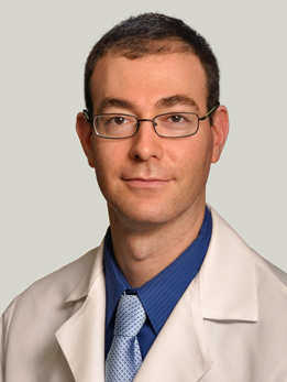
Dr. Horowitz is a skull base and neuro-oncology surgeon who also serves as Director of Quality and Associate Program Director for the Neurological Surgery residency. His laboratory research has identified novel genes driving meningioma and pediatric glioma formation, with work published in Nature Genetics and PNAS, and is funded by the DoD Neurofibromatosis Research Program. He holds a PhD in neuroscience from Northwestern and completed residency at Brigham and Women's/Boston Children's with a skull base fellowship at MD Anderson. Dr. Horowitz co-authored a comprehensive review of meningioma biology and management (Future Oncology, 2018) and routinely sees patients for both upfront surgery and second-opinion review of incidentally discovered meningiomas at UChicago.
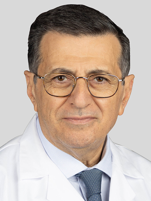
Dr. Comair is Section Chief of Neurosurgical Oncology and a pioneer of awake craniotomy for tumors in eloquent cortex. He has authored over 120 peer-reviewed publications and two neurosurgery textbooks, and he founded the first comprehensive epilepsy surgery program in the Middle East and North Africa. He trained at the Montreal Neurological Institute and previously held faculty positions at Cleveland Clinic, UCLA, Johns Hopkins, Baylor, and the American University of Beirut. Dr. Comair brings decades of experience in the microsurgical resection of supratentorial tumors — including convexity and parafalcine meningiomas overlying eloquent brain — where preserving function matters as much as removing the tumor.
What Is a Meningioma?
A meningioma is a tumor that grows from the meninges — the thin layers of tissue that wrap around your brain and spinal cord. Despite the name, meningiomas are not technically brain tumors: they arise from the lining outside the brain and press on the brain as they grow. That distinction matters, because it usually means a skilled surgeon can separate the tumor from healthy brain tissue rather than cutting into it.
Meningiomas are the most common primary tumor of the central nervous system, accounting for roughly 40% of all brain and spinal cord tumors in the United States. They're about twice as common in women as in men, and they most often appear between ages 40 and 70. Most are discovered either because they cause symptoms (headaches, seizures, weakness) or, increasingly, as an incidental finding on an MRI ordered for some other reason — a minor car accident, a migraine workup, a stroke evaluation.
The good news is that the overwhelming majority of meningiomas are benign (WHO grade 1). They grow slowly, they don't invade the brain, and when they can be fully removed, most patients are cured. This article focuses on the three most common and most surgically straightforward locations: convexity meningiomas (on the surface of the brain, under the skull), parasagittal meningiomas (alongside the large vein that runs down the middle of the skull), and falcine meningiomas (attached to the fold of dura between the two hemispheres). Skull base meningiomas — those at the bottom of the brain, near the cranial nerves and carotid arteries — are a separate, more technically demanding category and are covered in a different article.
At a Glance
- Meningiomas are the most common primary brain tumor — about 40% of all brain tumors — and the vast majority are benign (WHO grade 1)
- Many small, asymptomatic meningiomas can safely be watched with MRI rather than treated
- When surgery is needed, the goal is to remove the tumor along with its attachment to the dura (the lining of the brain) — this is measured by the Simpson grade
- Convexity, parasagittal, and falcine meningiomas are the most surgically accessible locations, and outcomes after complete removal are excellent
- Radiosurgery (a focused, single-dose radiation treatment) is an option for smaller tumors or residual tumor near critical structures
Have imaging or a diagnosis already?
We'll have a specialist review your MRI and records — often within 24 hours.
What Does a Meningioma Feel Like?
A lot of meningiomas feel like nothing at all. Because they grow slowly — often over years — the brain has time to accommodate them, and many are only picked up on an imaging scan ordered for an unrelated reason. When meningiomas do cause symptoms, those symptoms depend on exactly where the tumor is pressing.
Convexity meningiomas (over the brain's surface)
- New-onset seizures — often the very first sign, especially for tumors over the motor or sensory areas
- Gradual weakness or numbness on one side of the body
- Headaches, sometimes worse in the morning or with coughing and straining
- Personality changes or trouble with concentration, if the tumor is over the frontal lobe
- Changes in vision, language, or memory depending on which lobe is involved
Parasagittal and falcine meningiomas (near the midline)
- Weakness in one leg (or both legs, for falcine tumors near the motor strip) — because the part of the brain that controls the legs sits right next to the midline
- Seizures starting in a foot or leg
- Progressive difficulty walking
- Bladder urgency or incontinence, with large tumors compressing both frontal lobes
- Subtle apathy, slowed thinking, or loss of initiative — these are easily mistaken for depression or aging
If any of these symptoms are new, persistent, or getting worse, it's worth getting an MRI. Meningiomas are often treatable, and catching them before they cause permanent damage is far easier than repairing the damage after.
How Is a Meningioma Diagnosed?
The first step is imaging — almost always an MRI of the brain with and without contrast. Meningiomas have a very characteristic look on MRI: they sit outside the brain, they enhance brightly when contrast is given, and they usually show a dural tail — a streak of enhancement along the dura where the tumor is attached. That appearance alone is often enough for an experienced neuroradiologist or neurosurgeon to make the diagnosis with high confidence.
A CT scan is sometimes added, because it shows calcium and bone better than MRI does. Calcification inside a meningioma suggests the tumor has been there for a long time and is probably slow-growing. Bony changes in the overlying skull — either thickening (hyperostosis) or erosion — give the surgeon information about whether the bone itself needs to be removed and reconstructed.
A tissue diagnosis — the definitive answer about what the tumor actually is — requires surgery. Pathologists examine the removed tumor under the microscope and assign it a WHO grade (1, 2, or 3). More recently, molecular testing has started to play a role: certain genetic changes, like TERT promoter mutations or CDKN2A/B deletions, now automatically upgrade a meningioma to grade 3, because those changes predict a much higher risk of recurrence. For tumors that are going to be watched rather than operated on, imaging alone is what we follow.
Types of Meningioma: WHO Grades 1, 2, and 3
The World Health Organization groups meningiomas into three grades based on how they look under the microscope and — in the 2021 classification — on specific molecular changes. The grade is the single most important predictor of how the tumor will behave.
WHO Grade 1 — Benign (about 80% of meningiomas)
These are slow-growing, well-circumscribed tumors that don't invade the brain. When completely removed, most grade 1 meningiomas are cured. Common subtypes include meningothelial, fibrous, transitional, and psammomatous. Grade 1 tumors are the ones most often managed with either observation or a single operation.
WHO Grade 2 — Atypical (about 15-20%)
Atypical meningiomas grow faster, recur more often, and sometimes invade the brain itself. A tumor is classified as grade 2 if it shows increased cell division (4 or more mitoses per 10 high-power fields), brain invasion, or three or more of a set of specific microscopic features. The clear cell and chordoid subtypes are automatically grade 2. After surgery, most grade 2 meningiomas — especially those that aren't completely removed — are recommended for radiation therapy.
WHO Grade 3 — Anaplastic/Malignant (about 1-3%)
These are the rarest and most aggressive meningiomas. They behave like cancer: they invade the brain, grow quickly, recur reliably, and occasionally metastasize. A meningioma is grade 3 if it has very high mitotic activity (20+ mitoses per 10 high-power fields), frank malignant features, or specific molecular changes — TERT promoter mutations or CDKN2A/B homozygous deletions. These patients need aggressive surgery followed by radiation and close, lifelong follow-up.
How Is a Meningioma Treated?
Watch and wait — the right answer for many patients
One of the most important things to understand about meningiomas is that not every meningioma needs to be treated right away. A small, asymptomatic meningioma found incidentally on a scan can often be safely followed with serial MRIs — usually at 6 months, then yearly, then every 1-2 years if the tumor is stable. Many small meningiomas never grow meaningfully during a patient's lifetime. The decision to move from watching to treating is based on tumor growth, the development of symptoms, patient age and health, and the tumor's location.
Surgery — the definitive treatment when the tumor is symptomatic or growing
When a meningioma is causing symptoms, growing, or sitting in a location where growth would be dangerous, surgery is the first-line treatment. For convexity meningiomas, the operation is among the most gratifying in neurosurgery: the tumor is usually well-demarcated from the brain, the surgeon can remove it along with the attached dura and any involved bone, and recurrence is uncommon when the removal is complete.
Parasagittal and falcine meningiomas are trickier because they sit next to the superior sagittal sinus — the largest vein draining the top of the brain. If the tumor invades the sinus, the surgeon has to decide whether to leave a small amount of tumor behind (to preserve venous drainage and avoid a stroke) or to reconstruct the sinus. That judgment — when to push for complete resection and when to stop — is exactly what an experienced neurosurgical team is for.
The Simpson grade — how we measure how much was removed
Back in 1957, a neurosurgeon named Donald Simpson published a grading scale for meningioma resection that is still in use today. It captures not just whether the tumor was removed, but whether its dural attachment and any invaded bone were also taken.
- Simpson I — complete removal of the tumor, its dural attachment, and any invaded bone (the best result)
- Simpson II — complete removal plus coagulation (burning) of the dural attachment
- Simpson III — complete removal of the visible tumor, but the dura is left alone
- Simpson IV — partial removal
- Simpson V — biopsy only or decompression
Lower Simpson grades (I and II) are associated with lower recurrence rates. This is why the surgeon's approach to the dura — not just the tumor — matters so much for long-term outcome.
Radiosurgery — focused radiation for smaller or residual tumors
Stereotactic radiosurgery (Gamma Knife or CyberKnife) delivers a single, highly focused dose of radiation that kills tumor cells while sparing the surrounding brain. For meningiomas under about 3 cm that are in hard-to-reach locations, for patients who aren't surgical candidates, or for residual tumor left after surgery, radiosurgery is an excellent option. Long-term tumor control rates approach 90% for benign meningiomas treated this way.
Fractionated radiation therapy
For larger tumors or higher-grade (WHO 2 or 3) meningiomas, conventional fractionated radiation — delivered over several weeks — is often used after surgery. The NRG Oncology RTOG 0539 trial showed that adjuvant radiation after gross-total resection of a grade 2 meningioma produced 3-year progression-free survival over 93%.
What about chemotherapy?
There is no standard chemotherapy for meningioma. For tumors that keep coming back despite surgery and radiation, options include bevacizumab (an anti-angiogenic drug) and, increasingly, clinical trials of targeted therapies based on the tumor's specific molecular profile.
Considering surgery or planning a second opinion?
Our multidisciplinary team reviews complex cases together. You'll get a coordinated plan, not one opinion.
What Are the Outcomes?
For most people with a convexity, parasagittal, or falcine meningioma, the long-term outlook is very good. The main question isn't whether you'll survive the tumor — it's whether the tumor will come back and need a second treatment. Two things drive that: how completely the tumor was removed (the Simpson grade) and what the pathologist found under the microscope (the WHO grade).
Recurrence by Simpson grade (WHO grade 1 meningiomas)
| Simpson Grade | What it means | 5-year recurrence |
|---|---|---|
| Simpson I | Tumor, dura, and bone removed | ~5% |
| Simpson II | Tumor removed, dura coagulated | ~10% |
| Simpson III | Tumor removed, dura untreated | ~15-20% |
| Simpson IV | Partial removal | ~40% |
| Simpson V | Biopsy/decompression only | Nearly 100% progression |
Recurrence by WHO grade
| WHO Grade | 5-year recurrence | What to expect |
|---|---|---|
| Grade 1 (benign) | ~5-15% | Most are cured with complete removal |
| Grade 2 (atypical) | ~30-50% | Radiation often recommended after surgery |
| Grade 3 (anaplastic) | ~70-80% | Aggressive; needs surgery + radiation + close follow-up |
A few things to take away from these numbers. First, Simpson grade and WHO grade are not independent — a surgeon who can achieve a Simpson I resection on a grade 1 tumor has given that patient an excellent chance of cure. Second, the difference between a 5% and a 40% recurrence rate comes down to surgical judgment and technique. And third, even when recurrence happens, there's usually another line of treatment — re-operation, radiosurgery, or fractionated radiation. Choosing an experienced meningioma team at the start is what keeps the number of future treatments as small as possible.
References
Have Questions About Meningioma?
Our team is here to review your case, discuss your options, and help you take the next step.
Schedule: (773) 702-2123