Our Specialists for Mesial Temporal Sclerosis / Temporal Lobe Epilepsy
Deciding whether a temporal lobe is safe to operate on — and which operation to offer — depends on a detailed, multi-day workup and years of experience reading the results. At UChicago, the same functional neurosurgeons who run the epilepsy surgery program also perform laser ablations, stereo-EEG, and open resections, so your evaluation and your treatment come from one team.
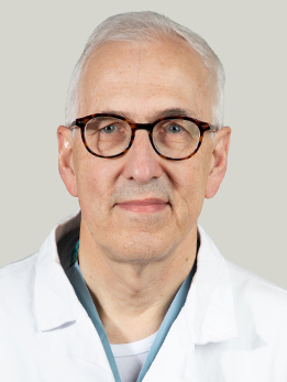
Dr. Warnke is an international leader in functional neurosurgery and has performed over 6,000 stereotactic surgeries and more than 3,000 brain tumor surgeries. He is only the second neurosurgeon worldwide to perform laser hemispherotomy, and he has completed over 400 laser ablation surgeries since arriving at UChicago. He is funded by four NIH grants including the BRAIN Initiative, and he directs the NAUTILUS trial for thalamic stimulation in drug-resistant epilepsy. Dr. Warnke runs the UChicago epilepsy surgery program and has personally performed more than 400 laser ablations since arriving at UChicago, including stereotactic laser amygdalohippocampectomy for mesial temporal sclerosis. If you are being evaluated for LITT or a stereo-EEG-guided resection at UChicago, he is likely your surgeon.
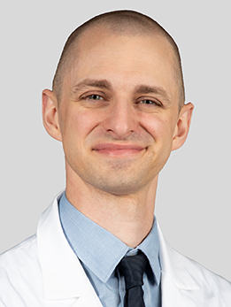
Dr. Satzer is a functional neurosurgeon specializing in epilepsy surgery, laser ablation, and deep brain stimulation. He is a recipient of the American Epilepsy Society Junior Investigator Award, and his research focuses on local field potentials and aperiodic neural activity as biomarkers for seizures and neuromodulation. His recent work has appeared in Brain Stimulation (2025). Dr. Satzer specializes in laser ablation, stereo-EEG, and open temporal lobe surgery for drug-resistant epilepsy, and has published on seizure and EEG prognostic factors after stereotactic laser amygdalohippocampectomy for mesial temporal lobe epilepsy (Frontiers in Neurology, 2021).
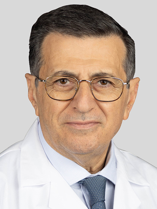
Dr. Comair is Section Chief of Neurosurgical Oncology and a pioneer of awake craniotomy for tumors in eloquent cortex. He has authored over 120 peer-reviewed publications and two neurosurgery textbooks, and he founded the first comprehensive epilepsy surgery program in the Middle East and North Africa. He trained at the Montreal Neurological Institute and previously held faculty positions at Cleveland Clinic, UCLA, Johns Hopkins, Baylor, and the American University of Beirut. Dr. Comair trained at the Montreal Neurological Institute and built the first comprehensive epilepsy surgery program in the Middle East; his published work on tailored temporal resections and tumor-associated epilepsy grew out of decades of high-volume temporal lobe surgery (Journal of Child Neurology, 1999).
What Is Temporal Lobe Epilepsy?
Temporal lobe epilepsy (TLE) is a type of focal epilepsy — meaning the seizures start in one specific area of the brain, rather than spreading across both sides at once. In TLE, that area is the temporal lobe, the part of the brain sitting above and behind your ear that handles memory, emotion, smell, and parts of language. TLE is the most common focal epilepsy in adults and, by a wide margin, the form of epilepsy most likely to be cured by surgery.
The majority of adults with TLE have a specific pathology called mesial temporal sclerosis (also known as hippocampal sclerosis). On MRI and under the microscope, the hippocampus — a seahorse-shaped structure tucked into the inner part of the temporal lobe — is shrunken and scarred, with loss of neurons in specific sub-regions. Many patients with mesial temporal sclerosis have a history of a childhood febrile seizure, head injury, or brain infection decades before their epilepsy begins, though we still cannot fully explain why some people develop sclerosis and others do not.
Not every temporal lobe seizure comes from the hippocampus. Some originate from a small tumor, a cortical malformation, a cavernoma, or an area of scar tissue elsewhere in the temporal lobe. Sorting out exactly where seizures are starting — and which structures can safely be removed — is the whole point of the presurgical workup.
At a Glance
- Temporal lobe epilepsy is the most common type of focal epilepsy in adults and the most common type successfully treated with surgery
- The usual underlying cause is mesial temporal sclerosis — scarring and cell loss in the hippocampus, the brain's memory structure
- If two well-chosen seizure medications have failed, your epilepsy is considered drug-resistant and surgical evaluation is recommended
- A randomized trial (Wiebe 2001) showed that 58-64% of surgical patients were seizure-free at one year, compared with 8% on medication alone
- Modern options range from traditional anterior temporal lobectomy to minimally invasive laser ablation (LITT) through a small hole in the skull
Have imaging or a diagnosis already?
We'll have a specialist review your MRI and records — often within 24 hours.
What Does It Feel Like?
Temporal lobe seizures rarely look like the dramatic, full-body convulsions people expect. Most are focal seizures with impaired awareness, which means the person stops what they are doing, stares blankly, and performs small, repetitive movements for a minute or two. Afterward, they are tired, confused, and often have no memory of what happened. Many patients learn about their seizures mainly from what their family describes.
The aura — a warning before the seizure
Most people with TLE feel a warning, called an aura, in the seconds before they lose awareness. Auras are themselves small seizures, and their character often points to the temporal lobe:
- A sudden rising feeling in the stomach, like the drop of a roller coaster
- An intense wave of fear, dread, or déjà vu (feeling that something has happened before)
- Jamais vu — the opposite, where a familiar place suddenly feels unfamiliar
- A strange smell or taste, often described as unpleasant or burning
- A sudden dreamy, distant feeling or a vivid memory flashing back
What happens during the seizure itself
- Staring and sudden unresponsiveness
- Automatisms — lip smacking, chewing, picking at clothing, or fumbling with objects
- A hand or arm that stiffens or posture that twists to one side
- Speech that stops or becomes garbled
- Occasional progression to a full-body (generalized tonic-clonic) convulsion
After the seizure
- Several minutes of confusion, slurred speech, and exhaustion
- Trouble finding words, especially after left temporal seizures
- No memory of the event itself
Over years, untreated temporal lobe seizures can also cause progressive memory problems, depression, and anxiety. This is one reason why early referral to an epilepsy center matters — the longer seizures continue, the more the surrounding memory networks are affected.
How Is It Diagnosed?
Diagnosing TLE and deciding whether surgery is an option requires several tests, usually done across a few visits at a specialized epilepsy center. The goal is to answer two questions: Where exactly are the seizures starting? and Can that area be removed safely without damaging memory, language, or vision?
Drug-resistant epilepsy — when to start thinking about surgery
The International League Against Epilepsy defines drug-resistant epilepsy as failure of two appropriately chosen, adequately dosed anti-seizure medications to control seizures. Once a patient hits that threshold, the chance that a third or fourth medication will work drops to around 5%. That is the point at which guidelines recommend referral to an epilepsy surgery center — not as a last resort, but as the next evidence-based step.
Phase I — the non-invasive workup
- High-resolution MRI using a dedicated epilepsy protocol, which looks specifically for hippocampal atrophy, abnormal signal, loss of internal structure, and subtle cortical malformations
- Video-EEG monitoring in the hospital for several days, where medications are reduced to capture seizures and confirm which side of the brain they are starting from
- PET scan, which often shows reduced metabolism in the affected temporal lobe between seizures
- Ictal SPECT, an imaging study where a tracer is injected during a seizure to map the blood flow changes as the seizure starts
- Neuropsychological testing — several hours of memory, language, and attention testing that establishes a baseline and helps predict how surgery will affect cognition
- Functional MRI and, for some patients, a Wada test, to determine which side of the brain controls language and memory
Phase II — stereo-EEG (intracranial recording)
When the non-invasive tests agree — for example, MRI shows left mesial temporal sclerosis, seizures on scalp EEG start on the left, PET is reduced on the left, and memory testing is worse for verbal (left-hemisphere) material — surgery can often be offered without further invasive testing. When the picture is murky, the next step is stereo-EEG (SEEG): thin recording electrodes are placed through tiny holes in the skull, under robotic guidance, directly into the suspected seizure zones. The patient spends several days in the hospital while we record actual seizures from inside the brain. SEEG is how we resolve the difficult cases — non-lesional TLE, bitemporal seizures, or suspected temporal-plus epilepsy where seizures spread to nearby regions.
Types of Temporal Lobe Surgery
Once the workup is complete and the seizure focus is clearly identified, there are three main surgical options. Your team will recommend one based on your anatomy, which side of the brain is involved, what the MRI shows, and how your memory is already functioning.
Anterior temporal lobectomy (ATL)
This is the classic, most-studied epilepsy operation. Through a standard craniotomy, the surgeon removes the front part of the temporal lobe — typically about 4-4.5 cm on the non-dominant (usually right) side and a little less on the dominant (usually left) side — together with the amygdala and the front portion of the hippocampus. ATL is the procedure used in the landmark Wiebe and Engel randomized trials, and it produces the highest published seizure-freedom rates for mesial temporal sclerosis: roughly 60-70% of patients are free of disabling seizures at one year, with durable results extending well beyond a decade.
Selective amygdalohippocampectomy (SAH)
Selective amygdalohippocampectomy is a more targeted open operation that removes only the amygdala and hippocampus while preserving the overlying temporal lobe cortex. It can be performed through a small trans-sylvian, sub-temporal, or trans-cortical corridor. The idea is to achieve seizure freedom with less impact on neocortical functions like language and naming. Meta-analyses suggest seizure-freedom rates are similar to ATL or slightly lower, with some evidence of better preserved naming and verbal memory on the dominant side.
Laser interstitial thermal therapy (LITT)
LITT — also called stereotactic laser amygdalohippocampectomy (SLAH) — is a minimally invasive alternative. Through a single 3.2-millimeter hole in the skull, a laser fiber is placed under MRI guidance along the length of the hippocampus. Inside the MRI scanner, heat is delivered under real-time thermal imaging, ablating the amygdala and hippocampus from within. There is no craniotomy, most patients go home within 24-48 hours, and neuropsychological studies suggest better preservation of naming and verbal memory compared with open surgery — at the cost of a modestly lower seizure-freedom rate (about 55-60% for mesial temporal sclerosis in large series). UChicago is one of the highest-volume LITT centers in the world.
Responsive neurostimulation and deep brain stimulation
For patients who are not candidates for resection or ablation — for example, those with seizures coming from both temporal lobes, or from eloquent cortex — implantable devices that detect and disrupt seizures (RNS) or continuously stimulate deep targets (DBS of the anterior nucleus of the thalamus) can substantially reduce seizure frequency, although they rarely produce complete seizure freedom.
How Is It Treated?
Medications come first
Everyone with new temporal lobe epilepsy starts with anti-seizure medications. Modern options — levetiracetam, lamotrigine, lacosamide, oxcarbazepine, brivaracetam, cenobamate, and others — control seizures in roughly two-thirds of patients. If one medication fails, a second is tried. If that one fails too, the chance of becoming seizure-free on a third medication is low, and it is time for a specialized epilepsy center to take over.
Why surgery, and why earlier rather than later
In 2001, Samuel Wiebe and colleagues published the first randomized trial of epilepsy surgery. Eighty patients with temporal lobe epilepsy were assigned to either surgery or continued medication. At one year, 58% of the surgical group was free of disabling seizures (64% of those who actually had surgery), compared with just 8% of the medication group. Quality of life and employment outcomes also favored surgery. A second randomized trial, ERSET (Engel 2012), confirmed the finding in patients with shorter seizure duration — zero of 23 medical patients and 11 of 15 surgical patients were seizure-free in the second year. Despite this evidence, surgery remains underutilized: the average patient waits more than 20 years from diagnosis to referral.
The day of surgery — open resection
For anterior temporal lobectomy or selective amygdalohippocampectomy, you are asleep under general anesthesia. The surgeon makes an incision behind the hairline, removes a small section of skull, and uses microsurgical technique, neuronavigation, and in some cases intraoperative ECoG (recording from the brain surface) to define and remove the epileptogenic tissue. The operation takes 3-5 hours. Most patients spend one night in the ICU and 3-4 nights total in the hospital.
The day of surgery — laser ablation
For LITT, a small stereotactic frame or robot is used to place the laser fiber through a single 3.2 mm skull opening. You are then moved into the MRI scanner, and the ablation is performed in 1-3 minute pulses with real-time thermal mapping of the target. There is no open exposure of the brain. Most patients go home within 24-48 hours and return to desk work within about a week.
After surgery
Anti-seizure medications are continued for at least the first year. Many patients are able to gradually taper and sometimes stop medications entirely if they remain seizure-free for 2-3 years, though this decision is individualized. Follow-up includes MRI, neuropsychological retesting, and regular visits with your epileptologist.
Considering surgery or planning a second opinion?
Our multidisciplinary team reviews complex cases together. You'll get a coordinated plan, not one opinion.
What Are the Outcomes?
The most widely used outcome measure in epilepsy surgery is the Engel classification, introduced by Jerome Engel in 1987. It groups patients into four classes based on how their seizures respond:
- Engel I — free of disabling seizures (the goal)
- Engel II — rare disabling seizures ("almost seizure-free")
- Engel III — worthwhile improvement
- Engel IV — no worthwhile improvement
For mesial temporal sclerosis treated with anterior temporal lobectomy, long-term studies consistently show that 60-70% of patients remain in Engel class I at one year, and roughly half remain seizure-free at 10 years. The numbers are best when MRI shows clear hippocampal sclerosis, seizures are clearly coming from a single side, and surgery is performed sooner rather than after decades of drug-resistant epilepsy.
References
Have Questions About Temporal Lobe Epilepsy?
Our team is here to review your case, discuss your options, and help you take the next step.
Schedule: (773) 702-2123