Our Specialists for Unruptured Intracranial Aneurysm
Deciding what to do about an unruptured aneurysm is as important as the procedure itself. Our cerebrovascular team reviews every case together — open microsurgeons, endovascular specialists, and neuroradiology — so you get a balanced recommendation rather than one tool's opinion of every problem.
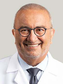
Dr. Awad is Section Chief of Vascular Neurosurgery and a world authority on cerebral cavernous malformations. He discovered the Common Hispanic CCM1 and Ashkenazi Jewish CCM2 mutations and leads the nation's first designated CCM Center of Excellence, with continuous NIH funding since 1998. He has authored more than 400 publications with over 100,000 citations, serves as past President of the Congress of Neurological Surgeons, and is an elected member of the Association of American Physicians. Dr. Awad leads UChicago's Neurovascular Surgery Program and has spent three decades operating on cerebral aneurysms and running the multicenter trials that shaped modern practice; if you are considering open microsurgical clipping for a complex unruptured aneurysm at UChicago, he is very likely your surgeon.
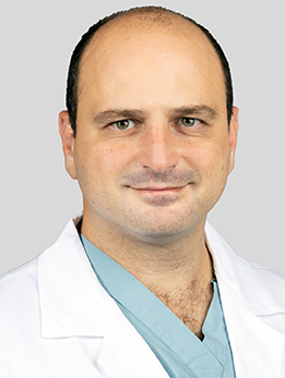
Dr. Doron is an endovascular neurosurgeon who brought more than 2,500 endovascular procedures with him from his Harvard/MGH fellowship. He performs both catheter-based and open microsurgical treatment of aneurysms, stroke, and vascular malformations, and he is building out thrombectomy capability across UChicago's regional network. He holds a PhD in biomedical engineering from Hebrew University of Jerusalem. Dr. Doron is a dual-trained open and endovascular neurosurgeon who performed more than 2,500 endovascular cases during his fellowship at Mass General, and has published technical refinements of Pipeline flow-diversion deployment for difficult aneurysm anatomy (Interventional Neuroradiology, 2023).
Dr. Zakaria is an endovascular neurosurgeon performing catheter-based treatment of stroke, brain aneurysms, and vascular malformations. He practices alongside the department's expanding cerebrovascular program, contributing to the regional thrombectomy and neurovascular service at UChicago. Dr. Zakaria sees unruptured aneurysm patients weekly at UChicago and has published on flow-diversion device selection for complex fusiform posterior-circulation aneurysms, an area where technical judgment matters as much as the device itself.
What Is an Unruptured Intracranial Aneurysm?
An intracranial aneurysm is a weakened, ballooned-out pouch on the wall of an artery inside your brain. Most look like small berries hanging off the side of a blood vessel where it branches (which is why doctors call them saccular or berry aneurysms). An unruptured aneurysm is one that hasn't bled — it's just sitting there, and you've learned about it because it showed up on a scan.
Unruptured aneurysms are more common than most people realize. Careful imaging studies suggest that roughly 2-3% of adults carry one without knowing it. The good news is that the large majority of these aneurysms will never rupture during a person's lifetime. The difficult news is that when a brain aneurysm does rupture, it causes a subarachnoid hemorrhage — a type of stroke that kills about a third of patients and disables many more. That's the tension at the heart of every decision about an unruptured aneurysm: a problem that is usually silent, but occasionally catastrophic.
Finding one is not an emergency. It is, however, an important conversation — and one that benefits from being had with a team that sees these every week.
At a Glance
- An unruptured aneurysm is a weak, ballooned-out spot on a brain artery — most are found by accident on a scan for something else.
- The majority of small aneurysms never bleed; the average annual rupture risk across all comers is under 1% per year.
- Risk depends on size, location, your age, blood pressure, smoking status, and family history — the PHASES score puts these together into a 5-year estimate.
- Three main treatment options exist: careful observation with repeat imaging, open microsurgical clipping, or endovascular repair (coiling, stent-assisted coiling, or flow diversion).
- Whether to treat — and how — is a judgment call that should be made at a center that does all three approaches in high volume.
Have imaging or a diagnosis already?
We'll have a specialist review your MRI and records — often within 24 hours.
What Does It Feel Like?
Here is the part that surprises most patients: unruptured aneurysms usually cause no symptoms at all. The vast majority are discovered incidentally — on an MRI ordered for migraines, a CT scan after a car accident, or a screening study because a family member had a brain bleed.
When unruptured aneurysms do cause symptoms
Only larger aneurysms, or aneurysms in specific locations, produce warning signs. These come from the aneurysm pressing on nearby nerves or brain tissue:
- A new, drooping eyelid with a dilated pupil on one side — classically from a posterior communicating artery aneurysm compressing the third cranial nerve. This is one of the few situations where an unruptured aneurysm demands urgent attention.
- Double vision or trouble moving one eye
- Facial pain or numbness on one side
- Visual changes — loss of peripheral vision from an aneurysm pressing on the optic nerve
- Persistent headaches in one spot — uncommon, and usually only with large or giant aneurysms
Warning signs that an aneurysm may have bled
These symptoms are different — they mean a ruptured aneurysm and they are a 911 emergency:
- The worst headache of your life, coming on in seconds ("thunderclap" headache)
- Sudden stiff neck, nausea, or vomiting with a severe headache
- Brief loss of consciousness or confusion
- Sudden weakness, numbness, or trouble speaking
If any of these happen, call 911 — do not wait for an appointment.
How Is It Diagnosed?
Most unruptured aneurysms are found on imaging done for an unrelated reason. Once an aneurysm is suspected, your team will want a more detailed look before making any decisions.
Imaging studies
CT angiography (CTA) and magnetic resonance angiography (MRA) are usually the first tests. Both give a 3-D picture of the arteries in your brain and can measure the aneurysm's size, shape, and location. They're non-invasive — no catheters, just a scan.
Digital subtraction angiography (DSA) — a catheter-based angiogram — is still the gold standard. A small catheter is threaded from the wrist or groin up into the arteries of the neck, and contrast dye is injected while a high-resolution camera films the blood flow. DSA shows the finest anatomic detail, which matters when the team is deciding exactly how to treat an aneurysm and which tools to use.
What the team is looking for
When we read your scans we are trying to answer very specific questions:
- How big is it? Size is the strongest single predictor of rupture.
- Where is it? Some locations — especially the posterior circulation — are riskier than others.
- What shape? Irregular, multilobed ("daughter sac") aneurysms carry a higher rupture risk than smooth round ones.
- Is it growing? If there's an older scan to compare, growth is a red flag.
- Are there others? About 20-30% of patients with one aneurysm have more than one.
Your team will also ask about your blood pressure, smoking history, family history of aneurysms or brain bleeds, and any prior subarachnoid hemorrhages — all of which feed directly into the risk calculation.
Types of Intracranial Aneurysms
Not all aneurysms behave the same way. The two features that matter most are shape and location, because they determine both the rupture risk and the best way to treat them.
By shape
- Saccular (berry) aneurysms are by far the most common. They look like a small balloon on the side of an artery, usually at a branch point where blood flow stresses the vessel wall. These are the typical aneurysms treated with clipping or coiling.
- Fusiform aneurysms are longer, spindle-shaped dilations involving a whole segment of the artery rather than a side pouch. They can't be clipped the same way — they usually need flow diversion or a bypass approach.
- Dissecting and mycotic aneurysms are less common and result from a tear in the artery wall or an infection; they require individualized treatment plans.
By location
- Anterior communicating artery (ACom) — the most common site for ruptured aneurysms. Located at the front of the brain where the two sides' blood supplies connect. Deep midline location makes these challenging for open surgery.
- Posterior communicating artery (PCom) — sit where the carotid gives off a branch to the back of the brain. A growing PCom aneurysm is one of the few that can cause an obvious symptom (a drooping eyelid with a dilated pupil).
- Middle cerebral artery (MCA) — often have multiple branches coming off the aneurysm itself, which traditionally has made them better candidates for microsurgical clipping than for coiling.
- Ophthalmic / paraclinoid internal carotid artery — sit near the optic nerve. Large ones can affect vision. These are increasingly treated with flow diversion.
- Basilar tip and other posterior circulation — at the back of the brain. These carry a higher rupture risk at any given size and are surgically demanding; many are now treated endovascularly.
How Is It Treated?
There are three legitimate options for an unruptured aneurysm: watch it, clip it, or fix it from the inside (coiling or flow diversion). The right choice depends on the aneurysm, your overall health, and your values. A good cerebrovascular team is one that can offer any of them and has no financial or technical reason to push you toward one.
Observation with surveillance imaging
For small, stable aneurysms in low-risk locations — especially in older patients — the safest thing is often to leave the aneurysm alone and watch it. You'll get repeat MRA or CTA imaging, typically at 6-12 months and then annually or every other year, looking for growth or shape change. Blood pressure control and stopping smoking are the two most important things you can do to lower your risk while you watch — smoking roughly doubles rupture risk, and quitting is the single most effective preventive intervention.
Microsurgical clipping
Clipping is the original aneurysm operation and remains the most durable cure. Through a small opening in the skull (a craniotomy), the surgeon gently parts the brain to expose the aneurysm and places a tiny titanium clip across its neck — like pinching off the base of a balloon — permanently closing it off from circulation. When complete, clipping has the lowest long-term recurrence rate of any treatment.
Clipping is often the preferred option for middle cerebral artery aneurysms, complex aneurysms with branches coming off them, wide-necked aneurysms in younger patients, and situations where endovascular access is unsuitable. At UChicago, clipping is performed with intraoperative angiography or indocyanine green video angiography to confirm the clip is placed perfectly before closing.
Endovascular coiling
Coiling is a minimally invasive alternative. A catheter is threaded from the wrist or groin up into the aneurysm itself, and soft platinum coils are packed inside the aneurysm sac until blood can no longer enter it. There is no craniotomy — most patients go home in a day or two.
Coiling is a strong option for many anterior circulation aneurysms, most posterior circulation aneurysms, and anyone whose medical condition makes open surgery risky. The trade-off is that coiled aneurysms can recanalize (the coils compact and blood starts entering the sac again) in about 10-20% of cases, so follow-up imaging is important, and a small number of patients need a second procedure.
Stent-assisted coiling
For aneurysms with a wide neck — where coils would fall out into the parent artery — a small stent is placed across the neck of the aneurysm first, acting as a scaffold so the coils stay where they belong. This requires you to take blood thinners (usually aspirin plus clopidogrel) for several months afterward.
Flow diversion (Pipeline and similar devices)
Flow diverters changed the game for large, wide-necked, or fusiform aneurysms, especially along the internal carotid artery. Instead of packing the aneurysm with coils, a braided metal mesh tube (a Pipeline Flex or similar device) is placed in the parent artery across the mouth of the aneurysm. Blood stops flowing into the sac, which gradually thromboses and shrinks over the following months. Studies of flow diversion for large carotid aneurysms show complete occlusion in about 75-85% of patients at one year, with a periprocedural major complication rate in the 5-6% range.
Flow diversion requires a blood-thinner regimen (dual antiplatelet therapy) for at least 6 months, so it isn't right for everyone — but for the aneurysms it fits, it's often the best option we have.
Putting it all together
In practice, the conversation goes something like this: How risky is your specific aneurysm if we leave it? How risky is each treatment, for you in particular? How many years of life do we expect to protect? For a 75-year-old with a 3 mm anterior circulation aneurysm, observation usually wins. For a 45-year-old smoker with a 9 mm basilar tip aneurysm, treatment almost always does. The in-between cases are where experience earns its keep.
Considering surgery or planning a second opinion?
Our multidisciplinary team reviews complex cases together. You'll get a coordinated plan, not one opinion.
What Are the Outcomes?
Outcomes fall into two categories: the natural history (what happens if you don't treat) and the periprocedural risk (what happens if you do). You're weighing one against the other, and both depend heavily on the specific aneurysm.
Natural history: how likely is rupture?
The overall annual rupture rate for unruptured aneurysms is often quoted as about 1% per year, but that single number hides a huge range. The PHASES score — derived from pooled data on more than 8,000 patients followed prospectively — gives a 5-year rupture estimate based on Population (ethnicity), Hypertension, Age, aneurysm Size, Earlier subarachnoid hemorrhage from another aneurysm, and Site.
| PHASES score | 5-year rupture risk | What it means |
|---|---|---|
| 0-2 | ~0.4% | Very low; observation usually safest |
| 3 | ~0.7% | Low |
| 4 | ~0.9% | Low |
| 5 | ~1.3% | Low-intermediate |
| 6 | ~1.7% | Intermediate; treatment often considered |
| 7 | ~2.4% | Intermediate-high |
| 8 | ~3.2% | High; treatment usually recommended |
| 9 | ~4.3% | High |
| ≥10 | ~5-17% | Very high; treatment strongly favored |
Two factors that are not in PHASES but matter anyway: smoking roughly doubles rupture risk, and a family history of aneurysmal subarachnoid hemorrhage in a first-degree relative raises it substantially. Both push the decision toward treatment for borderline cases.
Periprocedural risk: how safe is treatment?
Treatment is not free — and for small, low-risk aneurysms the risk of the procedure can exceed the risk of the aneurysm over 5 years. Large pooled analyses give us reasonable benchmarks at experienced centers:
- Microsurgical clipping of unruptured aneurysms: roughly 1.7% mortality and 6-7% overall morbidity, with better outcomes for anterior circulation and smaller aneurysms.
- Endovascular coiling of unruptured aneurysms: roughly 1-2% mortality and 4-5% unfavorable outcome, with risk rising for larger and posterior circulation aneurysms.
- Flow diversion for large or giant carotid aneurysms: 5-6% major stroke or neurologic death in the original trial, with complete occlusion rates around 75-95% at 1-5 years depending on aneurysm size and location.
Those numbers improve meaningfully at high-volume centers — which is why, when you're deciding where to have an aneurysm treated, volume and multidisciplinary review aren't marketing language; they're safety features.
References
Have Questions About an Unruptured Aneurysm?
Our team is here to review your case, discuss your options, and help you take the next step.
Schedule: (773) 702-2123