Our Specialists for Vestibular Schwannoma (Acoustic Neuroma)
Vestibular schwannomas sit in one of the most delicate places in the human body — the cerebellopontine angle, where the hearing nerve, balance nerve, and facial nerve run together into the brainstem. Experience and volume matter enormously here: both the decision of when to treat and the technique used to treat change hearing, facial function, and quality of life for decades afterward.
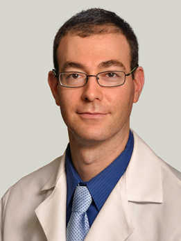
Dr. Horowitz is a skull base and neuro-oncology surgeon who also serves as Director of Quality and Associate Program Director for the Neurological Surgery residency. His laboratory research has identified novel genes driving meningioma and pediatric glioma formation, with work published in Nature Genetics and PNAS, and is funded by the DoD Neurofibromatosis Research Program. He holds a PhD in neuroscience from Northwestern and completed residency at Brigham and Women's/Boston Children's with a skull base fellowship at MD Anderson. Dr. Horowitz is a fellowship-trained skull base surgeon (MD Anderson) whose clinical practice is built around exactly this kind of cerebellopontine angle tumor; for most UChicago patients with a vestibular schwannoma that needs microsurgical removal, he is the primary surgeon of record.

Dr. Das is Director of Neurotrauma at UChicago's Level 1 Trauma Center and directs the Neurosurgical Trauma Fellowship, one of only a few dedicated neurosurgical trauma fellowships in the country. She manages both head trauma and acute spine pathology and has been named to the Bucksbaum-Siegler Institute for Clinical Excellence. She trained at the University of Minnesota for residency and completed a skull base fellowship at Cleveland Clinic. Dr. Das brings Cleveland Clinic skull base training and a focus on minimally invasive endoscopic and open corridors, and she partners with Dr. Horowitz on the multidisciplinary skull base team that evaluates every new vestibular schwannoma referral at UChicago.
Dr. Ali is Director of Endoscopic Neurosurgery at UChicago, specializing in minimally invasive keyhole approaches to skull base and intracranial lesions. His practice focuses on endoscopic techniques for pituitary and skull base tumors, using narrow corridors through the nose or small cranial openings to reach deep lesions with minimal disruption of surrounding tissue. Dr. Ali rounds out the skull base service with a practice focused on benign tumors of the cerebellopontine angle and a research interest in using augmented reality and advanced 3D visualization to plan safer approaches to the facial nerve during vestibular schwannoma surgery.
What Is a Vestibular Schwannoma?
A vestibular schwannoma is a slow-growing, benign tumor that arises from Schwann cells — the cells that form the insulating sheath around the vestibular portion of the eighth cranial nerve (the nerve of hearing and balance). It's sometimes called an acoustic neuroma, although that older name is a bit misleading since the tumor doesn't actually come from the hearing nerve itself.
These tumors typically grow inside the internal auditory canal — a narrow bony tunnel in the skull through which the hearing and balance nerves travel from the inner ear to the brainstem. As the tumor enlarges, it can push out into the cerebellopontine angle (CPA), a fluid-filled space where it can press on the facial nerve, the trigeminal nerve, the cerebellum, and eventually the brainstem itself.
Vestibular schwannomas are not cancer. They don't spread to other parts of the body, and they grow slowly — on average less than 1-2 millimeters per year. A small number don't grow at all, and a very small number even shrink on their own. Most are found in adults between 40 and 60 years old, and they are almost always on one side. Tumors on both sides are a hallmark of neurofibromatosis type 2 (NF2), a separate genetic condition that requires specialized care.
At a Glance
- A vestibular schwannoma is a benign (non-cancerous) tumor that grows from the insulating cells of the balance nerve (cranial nerve VIII)
- The most common first symptom is one-sided hearing loss, often with ringing (tinnitus) in that ear
- Many small tumors never need treatment — careful MRI surveillance is the right answer for most patients with small, stable tumors
- When treatment is needed, the three choices are stereotactic radiosurgery, microsurgical removal, or continued observation — the best choice depends on tumor size, your hearing, and your goals
- Facial nerve preservation and hearing preservation are the two outcomes that matter most, and both are strongly tied to surgical experience
Have imaging or a diagnosis already?
We'll have a specialist review your MRI and records — often within 24 hours.
What Does It Feel Like?
Because these tumors grow so slowly, symptoms usually come on gradually over months or years — which is part of why the diagnosis is often delayed. Many of these symptoms can be caused by more common conditions, but when they are one-sided and persistent, they deserve an ENT evaluation and an MRI.
The classic early symptoms
- One-sided hearing loss — often first noticed on the phone, or as trouble understanding speech in a noisy room. This is the most common presenting symptom.
- Tinnitus (ringing or buzzing) in one ear
- Imbalance or unsteadiness, especially in the dark or on uneven ground — true spinning vertigo is less common
- A feeling of fullness or pressure in one ear
When the tumor gets larger
- Numbness or tingling on one side of the face (from pressure on the trigeminal nerve)
- Facial weakness or twitching (less common — the facial nerve is surprisingly resilient)
- Headaches, especially in the back of the head
- Difficulty swallowing or hoarseness (from pressure on the lower cranial nerves)
- Unsteady walking or coordination problems (from pressure on the cerebellum)
A very large tumor can eventually compress the brainstem and cause hydrocephalus (buildup of spinal fluid inside the brain), which can become a medical emergency. This is uncommon today because most tumors are found long before they reach that size.
How Is It Diagnosed?
The workup usually starts with an audiogram — a formal hearing test — because most patients first notice one-sided hearing loss. Any asymmetric sensorineural hearing loss in an adult is a reason to image the inner ear and brainstem.
The gold standard test is a thin-slice MRI of the internal auditory canals with gadolinium contrast. Even tumors just a few millimeters across show up clearly on this study. Your team will measure the tumor in its largest dimension outside the internal auditory canal — this measurement drives both the Koos grade (see below) and most treatment decisions.
Additional testing may include:
- Repeat audiometry to quantify how serviceable your hearing is. The most common benchmark is the AAO-HNS 50/50 rule — a pure-tone average better than 50 dB and word recognition better than 50% is considered "serviceable."
- Balance testing (VNG or rotary chair) in some cases, to understand how well the opposite ear is compensating
- Facial nerve function grading using the House-Brackmann scale, which runs from I (perfectly normal) to VI (complete paralysis). This is your baseline for comparison after any treatment.
- Genetic testing for NF2 if you are young, have tumors on both sides, or have a family history
Unlike malignant tumors, a vestibular schwannoma almost never needs a biopsy — the MRI appearance is characteristic enough that your team can plan treatment directly from imaging.
Koos Grades: Size Matters
The Koos grading scale is the most widely used way to describe how big a vestibular schwannoma is and how much of its neighborhood it's involving. Treatment decisions — observation, radiosurgery, or microsurgery — map closely onto these grades.
Grade I — Intracanalicular
- The tumor sits entirely inside the internal auditory canal. No extension into the cerebellopontine angle.
- Usually the smallest and most likely to be watched safely. Many patients with Koos I tumors never need treatment.
Grade II — Small extension into the cerebellopontine angle
- The tumor extends out of the canal into the CPA but does not touch the brainstem. Typically less than 2 cm in extrameatal diameter.
- Good candidate for either observation, radiosurgery, or hearing-preserving microsurgery depending on hearing and patient preference.
Grade III — Touches the brainstem, no compression
- The tumor now reaches the brainstem but does not deform it. Usually 2-3 cm in extrameatal diameter.
- Often treated with radiosurgery or microsurgery. Pure observation is less commonly chosen.
Grade IV — Compresses and displaces the brainstem
- The largest tumors. They push the brainstem out of position and can obstruct the flow of spinal fluid.
- Microsurgical resection is usually the first choice — these tumors are generally too large for radiosurgery alone, and the pressure on the brainstem needs to be relieved.
How Is It Treated?
There is no single "right" treatment for vestibular schwannoma — there are three reasonable options, and the best one depends on your tumor's size, your hearing, your age, and what matters most to you. A dedicated skull base team will walk through all three with you.
1. Observation ("wait and scan")
For small, newly-diagnosed tumors — especially Koos I and II — simply watching with serial MRIs is often the right answer. Large natural-history studies show that many small tumors grow slowly or not at all. A recent multi-institutional volumetric study of nearly 1,000 observed patients found that the five-year growth-free survival was roughly 20%, but importantly, tumor growth on surveillance does not necessarily mean hearing will worsen faster, and many patients remain stable for years.
Observation usually means an MRI at 6 months, then yearly, then spacing out if stable. Treatment is recommended if the tumor grows meaningfully, if hearing declines quickly, or if you develop new symptoms.
2. Stereotactic radiosurgery (SRS)
Stereotactic radiosurgery — most commonly delivered with Gamma Knife or CyberKnife — focuses a single, precisely-targeted dose of radiation on the tumor in a one-day outpatient treatment. It does not remove the tumor; it stops it from growing. Long-term tumor control rates are excellent: roughly 94-97% at 5 and 10 years for tumors up to about 3 cm in diameter.
SRS is an especially good option for:
- Tumors under about 2.5-3 cm
- Older patients, or those who aren't good candidates for general anesthesia
- Tumors that have grown on surveillance
- Residual tumor after a previous subtotal microsurgical resection
Modern low-dose protocols (around 12-13 Gy to the tumor margin) have made facial nerve and trigeminal nerve complications rare. Hearing preservation in the short term is good, but hearing tends to decline over the decade following SRS, even when the tumor itself remains controlled.
3. Microsurgical resection
For larger tumors (especially Koos IV), for younger patients who want the tumor gone rather than irradiated, and for tumors that have failed radiosurgery, microsurgery is the definitive option. There are three standard skull base approaches, and the choice depends on tumor size, tumor location, and whether your hearing is worth preserving:
- Retrosigmoid approach — Behind the ear, through the back of the skull. The most flexible corridor, usable for any tumor size. Preserves the option of hearing preservation when hearing is still serviceable.
- Translabyrinthine approach — Through the mastoid bone and the semicircular canals. Sacrifices residual hearing on that side by design, but gives an unobstructed view of the facial nerve at the brainstem. Used for larger tumors when useful hearing has already been lost.
- Middle fossa approach — From above, through the floor of the temporal bone. Reserved for small, mostly intracanalicular tumors when hearing preservation is the main goal.
Every modern microsurgical resection uses intraoperative facial nerve monitoring and, when hearing preservation is the goal, auditory brainstem response monitoring. The CNS guidelines recommend these as standard of care. The goal is always maximal safe removal — and in select cases, the right answer is a planned subtotal resection to preserve facial function, with the remnant treated later by radiosurgery if needed.
What about medication?
There is no FDA-approved drug for sporadic vestibular schwannoma. Bevacizumab (an anti-VEGF antibody) is used in NF2-related bilateral tumors to slow growth and stabilize hearing, but it is not part of routine care for single sporadic tumors.
Considering surgery or planning a second opinion?
Our multidisciplinary team reviews complex cases together. You'll get a coordinated plan, not one opinion.
What Are the Outcomes?
For most people, a vestibular schwannoma is a once-in-a-lifetime diagnosis that does not shorten their life. The harder questions are about hearing and facial nerve function — and both are tied tightly to tumor size at the time of treatment and the experience of the team.
Tumor control
Long-term tumor control after stereotactic radiosurgery is approximately 97% at 5 years and 94% at 10 years for small-to-medium tumors. Complete microsurgical removal provides roughly 95-99% long-term control. Subtotal resection has a higher recurrence rate, which is why residual tumor is often followed closely or treated with radiosurgery.
Hearing preservation by approach and size
Hearing preservation is the outcome that varies most between treatments. The table below summarizes pooled estimates from recent meta-analyses and large surgical series — your individual odds depend on tumor size, preoperative hearing, and the experience of your team.
| Treatment | Serviceable hearing preserved (short term) | Notes |
|---|---|---|
| Observation | ~50-70% at 5 years | Depends on starting hearing and tumor growth |
| Gamma Knife SRS | ~55-75% early, <25% at 10 years | Hearing declines gradually after radiosurgery |
| Middle fossa (small tumor) | ~60-75% | Best for intracanalicular tumors <1.5 cm |
| Retrosigmoid (small-medium) | ~40-60% | Declines as tumor size grows beyond 1.5-2 cm |
| Translabyrinthine | 0% by design | Best facial nerve exposure; used when hearing is already gone |
Facial nerve preservation
In experienced microsurgical series, roughly 85-90% of patients retain good facial nerve function (House-Brackmann grade I or II) at long-term follow-up. For small tumors (<2 cm), that number approaches 95%. For very large Koos IV tumors, the risk of some degree of permanent weakness is higher, which is why subtotal resection is sometimes the smarter choice. After radiosurgery, permanent facial weakness is uncommon — well under 5% in modern low-dose protocols.
The experience effect
Across almost every published series, higher case volume predicts better outcomes — fewer complications, better facial nerve function, and a higher chance of hearing preservation. A dedicated, multidisciplinary skull base team that sees these tumors regularly will give you not only a better operation but also a more honest answer about whether you need one at all.
References
Have Questions About Vestibular Schwannoma?
Our team is here to review your case, discuss your options, and help you take the next step.
Schedule: (773) 702-2123